
10 Diagnose Pneumonia Truths Deadly Myths vs Fast Recovery
6 Life-Saving Steps to Diagnose Pneumonia Early — And the Tests That Confirm It
A 72-year-old man is brought to the emergency department with two days of confusion and reduced oral intake. He has a background of chronic obstructive pulmonary disease (COPD) and hypertension. His temperature is only 37.8°C. His cough is mild. The initial chest X-ray is reported as “possible right basal shadowing.” His white cell count is 11,800/mm³ — mildly raised, nothing dramatic. It would be easy to conclude that this is nothing serious, perhaps a viral illness or simple dehydration. Yet the real diagnostic question has only just begun: what evidence can separate community-acquired pneumonia from aspiration pneumonitis, pulmonary embolism, decompensated heart failure, urinary tract sepsis, or a dozen other conditions that can mimic lower respiratory tract infection? The answer lies not in a single test, but in a disciplined diagnostic process that integrates history, examination, laboratory data, imaging, and careful clinical reasoning.
This is a hypothetical educational case, but it reflects a daily reality in clinical practice. Pneumonia remains one of the leading infectious causes of death worldwide, yet its diagnosis is rarely as simple as a single abnormal chest X-ray. The consequences of both overdiagnosis and underdiagnosis are substantial: unnecessary antibiotics on one hand, and preventable respiratory failure, sepsis, and death on the other. Accurate diagnosis requires a systematic approach that integrates clinical history, physical examination, laboratory testing, imaging, and, when appropriate, microbiological confirmation.
A normal chest X-ray does not always mean the disease is absent.  Diagnosis is a process, not a single image on a screen.
This article provides a comprehensive, evidence-based guide to Pneumonia Diagnosis, written for medical students, residents, healthcare professionals, and informed readers who want to understand how clinicians separate true pneumonia from the conditions that mimic it. If you want to understand why laboratory values and imaging findings can sometimes be misleading, our detailed guide to medical diagnosis explained in 15 powerful steps explains how clinicians interpret these results in context.
Key Diagnostic Facts About Pneumonia
The following table summarizes the most clinically important facts about pneumonia diagnosis. This is not a substitute for full clinical evaluation, but it provides a rapid reference for healthcare professionals and students.
| Diagnostic Feature | Key Fact |
|---|---|
| Disease | Pneumonia (community-acquired, hospital-acquired, and ventilator-associated forms exist; this article focuses primarily on community-acquired pneumonia) |
| Main clinical suspicion | Acute onset of cough, fever, dyspnoea, and pleuritic chest pain; tachypnoea and focal chest signs on auscultation |
| Most important symptoms | Productive cough, fever or hypothermia, dyspnoea, pleuritic chest pain, fatigue, confusion (especially in older adults) |
| Important risk factors | Age ≥65 years, chronic lung disease, smoking, immunosuppression, recent influenza, aspiration risk, alcohol use disorder |
| First clinical step | History and physical examination to establish clinical suspicion and assess severity |
| Initial investigations | Pulse oximetry, chest X-ray (or lung ultrasound where expertise exists), complete blood count, basic metabolic panel |
| Important laboratory tests | Complete blood count with differential, blood urea nitrogen, electrolytes, C-reactive protein, procalcitonin (selected patients), blood cultures (moderate-to-severe disease) |
| Important imaging | Chest X-ray is the standard initial imaging modality; lung ultrasound is an accepted alternative in centres with appropriate expertise; CT is reserved for complicated or indeterminate cases |
| Confirmatory test | There is no single confirmatory test for all pneumonia. Diagnosis is clinical plus radiographic. Microbiological confirmation (sputum culture, blood culture, urinary antigen, PCR) identifies the pathogen when indicated |
| Important differential diagnoses | Acute bronchitis, pulmonary embolism, heart failure with pulmonary oedema, lung cancer, aspiration pneumonitis, COVID-19, tuberculosis |
| Major red flags | Respiratory rate ≥30/min, oxygen saturation <92% on room air, confusion, hypotension, multilobar infiltrates, need for mechanical ventilation |
| Specialist referral | Pulmonology or infectious diseases consultation for severe community-acquired pneumonia, suspected unusual pathogens, treatment failure, or immunocompromised patients |
| Diagnostic challenge | Distinguishing bacterial from viral pneumonia is frequently not possible on clinical, laboratory, or radiographic grounds alone; pretest probability and clinical context guide testing and treatment decisions |
This table is a snapshot. Every parameter in it will be expanded in the dedicated sections below.
Step 1 — When Should Doctors Suspect Pneumonia?
Typical Presentation: Pneumonia should enter the differential diagnosis when a patient presents with an acute or subacute respiratory illness accompanied by systemic features of infection. The classic presentation in adults includes a cough that may be productive of purulent sputum, fever with or without rigors, pleuritic chest pain, and breathlessness. Tachypnoea is often present and is a more sensitive indicator of lower respiratory tract infection than fever alone. Auscultation may reveal crackles, bronchial breath sounds, or dullness to percussion over the affected area, though physical examination findings can be subtle or absent, particularly in early disease or in older adults.
Early Symptoms: Early symptoms can be nonspecific. Fatigue, malaise, anorexia, and low-grade fever may precede respiratory symptoms by 24 to 48 hours. In some patients, particularly older adults, confusion or a decline in functional status may be the presenting complaint, without prominent fever or cough. This atypical presentation is a well-recognised diagnostic pitfall and is a common reason for delayed diagnosis.
Characteristic Features That Increase Clinical Suspicion: These include a focal area of crackles or bronchial breathing on auscultation, new hypoxaemia, tachypnoea, and a compatible radiographic opacity. However, none of these findings is perfectly sensitive or specific. The absence of crackles does not exclude pneumonia. A normal chest X-ray in the first 24 hours does not exclude pneumonia in a dehydrated or neutropenic patient.
Atypical Presentation: Atypical presentations are common in older adults, immunocompromised patients, and those with underlying chronic lung disease. In these groups, pneumonia may present with worsening confusion, falls, or decompensation of a comorbid condition rather than with classic respiratory symptoms. In children under 5 years of age, the World Health Organization defines pneumonia clinically by the presence of cough and/or difficulty breathing with fast breathing or lower chest wall indrawing — imaging is not required to make the diagnosis.
Duration and Course: Community-acquired pneumonia typically develops over hours to days. A more insidious course over weeks should raise suspicion for tuberculosis, fungal infection, or malignancy. Aspiration pneumonia may follow a witnessed aspiration event or occur in patients with dysphagia, altered consciousness, or neurological disease.
Epidemiological Clues: Recent influenza or COVID-19 infection increases the risk of secondary bacterial pneumonia. Exposure to contaminated water systems or recent hotel stays may suggest Legionella. Exposure to birds may suggest psittacosis. Travel to endemic areas may suggest tuberculosis or endemic fungal infections. These clues do not replace systematic evaluation, but they can meaningfully alter the pretest probability of specific pathogens.
Nonspecific Findings: Fatigue, myalgia, headache, and general malaise are common in both pneumonia and non-pneumonia conditions. If you want to understand how another major risk factor — diabetes — can quietly damage blood vessels and impair immune defence for years before producing symptoms, our detailed guide to diabetes blood sugar clues and early signs explains the diagnostic overlap in a clear, practical way.
Step 2 — Medical History 
The medical history is not a formality. It is the first and most important diagnostic instrument. The information gathered during history-taking determines which tests are ordered, how results are interpreted, and what alternative diagnoses are considered. A focused but thorough history should elicit the following:
- Presenting complaint: What exactly happened? The patient’s own words should be recorded, followed by a structured characterisation of each symptom.
- Onset and duration: Acute onset over hours suggests bacterial aetiology or pulmonary embolism. Subacute onset over days is typical of viral pneumonia. A more chronic course over weeks suggests tuberculosis or malignancy.
- Progression and severity: Is the breathlessness worsening? Can the patient complete a sentence without pausing? Are they able to maintain oral intake? These simple questions provide rapid severity assessment.
- Associated symptoms: Sputum colour and volume, haemoptysis, night sweats, weight loss, pleuritic pain, and extrapulmonary symptoms such as diarrhoea (which may accompany Legionella) or rash should be documented.
- Previous episodes and diagnoses: Recurrent pneumonia in the same lobe should raise suspicion of an underlying structural abnormality, foreign body, or malignancy.
- Medications and allergies: Immunosuppressive medications, proton pump inhibitors, and recent antibiotic exposure all influence the likely pathogens and the interpretation of culture results.
- Family history: Relevant for inherited immunodeficiency, cystic fibrosis, and alpha-1 antitrypsin deficiency.
- Social and occupational history: Smoking history, alcohol use, occupational exposure to dusts or fumes, and living arrangements (for example, nursing home residence) are all relevant.
- Travel and environmental exposure: Recent travel, hotel stays, exposure to air conditioning systems, hot tubs, or farm animals can point toward specific pathogens such as Legionella, Q fever, or endemic fungi.
- Immunisation history: Influenza, pneumococcal, COVID-19, and Haemophilus influenzae type b vaccination status should be recorded.
- Functional status: The patient’s ability to perform activities of daily living, cognitive baseline, and swallowing function are critical for assessing aspiration risk and determining appropriate site of care.
The history is a hypothesis-generating exercise. A patient who describes three days of productive cough and fever after a recent influenza-like illness is a different diagnostic problem from a patient who developed breathlessness after a period of immobility following surgery. If you want to understand how hypertension — a major risk factor for both cardiovascular and respiratory complications — is diagnosed and monitored, our detailed guide to hypertension diagnosis explained with BP readings and home monitoring explains the diagnostic criteria in a clear, practical way.
Physical Examination
Vital Signs and General Appearance: Tachypnoea (respiratory rate greater than 24 per minute in adults) is one of the most sensitive clinical signs of pneumonia. Tachycardia, fever or hypothermia, and hypotension all have prognostic significance. Pulse oximetry is mandatory and should be recorded on room air where possible. An oxygen saturation below 92% on room air in a patient without chronic lung disease is a red flag. The patient’s work of breathing, ability to speak in full sentences, mental status, and hydration status should be assessed immediately. Confusion in an older adult with a respiratory complaint is a significant warning sign.
For online earning ideas, freelancing, affiliate marketing, YouTube, Facebook monetization, and AI tips, visit ssthem.xyz for online earning and WhatsApp group links
Chest Examination: On inspection, look for asymmetry of chest movement, use of accessory muscles, and cyanosis. On palpation, reduced chest expansion and increased tactile fremitus over an area of consolidation may be detected, though these findings are operator-dependent. Dullness to percussion over a consolidated area is a classic but relatively insensitive sign. On auscultation, crackles (rales), bronchial breath sounds, and egophony are the most commonly taught signs of consolidation. However, their sensitivity is modest. In a systematic review, the presence of any abnormal auscultatory finding had limited diagnostic accuracy when used in isolation. The absence of crackles does not exclude pneumonia.
Findings Suggesting Complications: Pleural effusion may present with stony dullness, reduced breath sounds, and reduced vocal resonance. Sepsis may present with tachycardia, hypotension, altered mental status, mottled skin, and prolonged capillary refill. Respiratory failure may present with central cyanosis, inability to speak, use of accessory muscles, and paradoxical abdominal breathing.
Findings Suggesting Alternative Diagnoses: Unilateral leg swelling and pleuritic pain suggest pulmonary embolism. Bilateral basal crackles, raised jugular venous pressure, and peripheral oedema suggest heart failure. Cachexia, haemoptysis, and chronic cough suggest lung cancer or tuberculosis. Diffuse wheeze and prolonged expiration suggest asthma or COPD exacerbation.
Red Flags That Should Not Be Ignored
The following table summarizes clinically meaningful warning signs that require urgent attention. These are not exaggerated risks but genuine indicators of potentially serious underlying pathology.
| Red Flag | Why It Matters | Possible Concern | Appropriate Action |
|---|---|---|---|
| Respiratory rate ≥30/min | Indicates severe respiratory compromise | Severe pneumonia, impending respiratory failure | Urgent medical assessment; consider hospital admission and ICU evaluation |
| Oxygen saturation <92% on room air | Suggests significant gas exchange abnormality | Severe pneumonia, ARDS, pulmonary embolism | Supplemental oxygen; urgent hospital assessment |
| Confusion or altered mental status | May indicate hypoxaemia, sepsis, or central nervous system involvement | Severe pneumonia, sepsis, meningitis | Immediate medical evaluation; assess for sepsis |
| Systolic blood pressure <90 mmHg | Indicates haemodynamic compromise | Septic shock | Emergency resuscitation; ICU consideration |
| Multilobar infiltrates on imaging | Associated with increased mortality | Severe pneumonia | Hospital admission; consider ICU evaluation |
| Need for mechanical ventilation | Definitive major criterion for severe CAP | Respiratory failure | ICU admission |
| Temperature <36°C | Hypothermia is a poor prognostic sign in sepsis | Severe infection, sepsis | Urgent evaluation; blood cultures; broad-spectrum antibiotics |
| Rapidly progressive symptoms over hours | Suggests fulminant infection or alternative diagnosis | Necrotising pneumonia, pulmonary embolism, ARDS | Emergency assessment |
Differential Diagnosis
Pneumonia shares clinical features with several conditions. The ability to distinguish these is a core clinical skill. The clinical reasoning behind these distinctions is not simply pattern recognition. It is about integrating the time course, the patient’s risk profile, the physical examination findings, and the initial investigation results.
| Differential Diagnosis | Similar Features | Distinguishing Features | Key Investigation |
|---|---|---|---|
| Acute bronchitis | Cough, sputum production, low-grade fever | Usually no focal chest signs; chest X-ray is normal; systemic features are milder | Chest X-ray; clinical course |
| Pulmonary embolism | Dyspnoea, pleuritic chest pain, tachycardia, hypoxaemia | Sudden onset; risk factors (immobility, malignancy, oral contraceptives); clear chest on auscultation | CT pulmonary angiography; D-dimer (in low pretest probability) |
| Heart failure with pulmonary oedema | Dyspnoea, crackles, hypoxaemia | Orthopnoea, paroxysmal nocturnal dyspnoea, raised JVP, peripheral oedema; bilateral findings | Echocardiography; BNP/NT-proBNP; chest X-ray showing cardiomegaly and pleural effusions |
| Lung cancer | Cough, haemoptysis, weight loss, recurrent pneumonia | More chronic course; non-resolving pneumonia in the same lobe; smoking history | CT chest; bronchoscopy; biopsy |
| Aspiration pneumonitis | Cough, dyspnoea, hypoxaemia following aspiration event | Chemical injury rather than infection; often resolves without antibiotics; may progress to aspiration pneumonia | Clinical history; chest X-ray; response to supportive care |
| COVID-19 pneumonia | Fever, cough, dyspnoea, bilateral infiltrates | Viral aetiology; characteristic ground-glass opacities on CT; positive SARS-CoV-2 test | SARS-CoV-2 PCR or antigen test |
| Tuberculosis | Chronic cough, fever, night sweats, weight loss, haemoptysis | Subacute or chronic course; upper lobe infiltrates; cavitation | Sputum AFB smear and culture; nucleic acid amplification testing |
A young patient with sudden-onset pleuritic pain and a clear chest X-ray is more likely to have a pulmonary embolism than pneumonia. An older patient with orthopnoea, bilateral crackles, and a raised JVP is more likely to have heart failure. The diagnostic process should remain open to revision as new information emerges. It is worth remembering that respiratory and cardiac emergencies often overlap. If you have ever wondered how a heart attack differs from a cardiac arrest — and why the distinction matters in an emergency — our article on heart attack versus cardiac arrest explains the critical differences and the minutes that save a heart.
Diagnostic Tests
1. Chest X-ray: 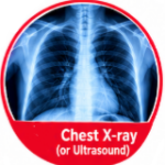 Ordered as the standard initial imaging investigation for suspected pneumonia. It is widely available, rapid, and provides information about the presence, location, and extent of pulmonary infiltrates. The classic finding is a new area of consolidation — an area of increased opacity that may contain air bronchograms. Lobar consolidation is characteristic of bacterial pneumonia, particularly Streptococcus pneumoniae. Interstitial patterns are more typical of viral or atypical pathogens. Multilobar involvement suggests severe disease. In adults presenting to hospital with suspected community-acquired pneumonia, NICE recommends that chest X-ray be performed within 4 hours of presentation, with antibiotic treatment initiated if pneumonia is confirmed. In patients with underlying lung disease, comparison with previous imaging is essential to determine whether the opacity is new. Chest X-ray has limited sensitivity in early disease, in dehydrated patients, and in neutropenic patients. It cannot reliably distinguish between bacterial and viral aetiology. Other causes of pulmonary opacity — pulmonary oedema, pulmonary infarction, atelectasis, malignancy, and ARDS — can mimic pneumonia. Early pneumonia, dehydration, and neutropenia can all produce a normal chest X-ray despite significant infection. A radiographic opacity alone does not establish the diagnosis of pneumonia; it must be accompanied by compatible clinical features.
2. Lung Ultrasound:  Increasingly used as a point-of-care diagnostic tool for pneumonia, particularly in emergency departments and intensive care units. It is radiation-free, repeatable, and can be performed at the bedside. The primary finding is subpleural consolidation, which appears as a tissue-like or anechoic area with blurred, irregular margins. Focal B-lines and reduced lung sliding may also be present. A meta-analysis using CT as the criterion standard showed that ultrasonography was more accurate than chest radiography at diagnosing community-acquired pneumonia. The 2025 American Thoracic Society (ATS) guidelines state that for adults with suspected community-acquired pneumonia, lung ultrasound is an acceptable diagnostic alternative to chest radiography in medical centres where appropriate clinical expertise exists. NICE also recognises that lung ultrasound can be used in the diagnosis of pneumonia in hospital. Lung ultrasound is operator-dependent. It cannot visualise the entire lung, and central lesions may be missed. It does not replace chest X-ray for the detection of complications such as pneumothorax or for the evaluation of mediastinal structures.
3. Complete Blood Count (CBC): A routine initial investigation in suspected pneumonia. It provides information about the white blood cell response and can identify leukocytosis, leukopenia, and thrombocytopenia. Leukocytosis (white blood cell count greater than 12,000/mm³) is common in bacterial pneumonia. Leukopenia (white blood cell count less than 4,000/mm³) is a poor prognostic sign and may be seen in severe infection or viral pneumonia. Thrombocytopenia (platelet count less than 100,000/µL) is a minor criterion for severe community-acquired pneumonia. The CBC is nonspecific. Leukocytosis can occur in many inflammatory conditions, and normal values do not exclude pneumonia. In older adults and immunocompromised patients, the white blood cell response may be blunted.
4. Inflammatory Biomarkers — CRP and Procalcitonin: 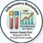 C-reactive protein (CRP) is an acute-phase reactant that rises in response to inflammation and infection. It is not specific for pneumonia and can be elevated in many other conditions. However, a very high CRP (greater than 100 mg/L) may increase the likelihood of bacterial infection. Procalcitonin (PCT) is a precursor of calcitonin that rises more specifically in response to bacterial infection. The 2016 and 2019 IDSA/ATS guidelines recommend cautious use of PCT and CRP in antibiotic decision-making due to variability in test sensitivity and the lack of consistent positive thresholds. A PCT level below 0.25 ng/mL suggests that bacterial infection is unlikely, and antibiotics may not be necessary in non-critically ill patients. A PCT level above 0.5 ng/mL is more suggestive of bacterial infection, though the threshold varies by clinical context. PCT is more useful for guiding the duration of antibiotic therapy than for establishing the initial diagnosis. PCT can be elevated in non-bacterial conditions and can be falsely low in early bacterial infection. It should never be used as the sole basis for withholding antibiotics in a patient with a high pretest probability of bacterial pneumonia.
5. Blood Cultures, Sputum Culture, and Urinary Antigen Tests: Blood cultures are recommended in patients with moderate-to-severe community-acquired pneumonia who require hospitalisation, particularly those with severe disease, immunocompromise, or suspected bacteraemia. They are positive in approximately 5–7% of patients with community-acquired pneumonia. Sputum culture can identify the causative pathogen and its antibiotic susceptibility profile. It has the highest microbiological yield of any non-invasive test when an adequate specimen is obtained, but it is performed in less than a quarter of patients in many clinical settings. The proportion of false-negative results ranges from 22% to 44% for common pathogens. Pneumococcal urinary antigen test detects the C-polysaccharide antigen of Streptococcus pneumoniae. Sensitivity is approximately 70–80%, and specificity is high. It can remain positive for days after infection and is not affected by prior antibiotic use. Legionella urinary antigen test detects Legionella pneumophila serogroup 1. Sensitivity is 70–100%, and specificity is 95–100%. It is particularly useful in severe community-acquired pneumonia, in patients with recent travel or hotel stays, and during outbreaks.
Laboratory Tests Table
| Test | Purpose | Possible Result | Interpretation | Limitations |
|---|---|---|---|---|
| Chest X-ray | Detect pulmonary infiltrates | New focal opacity | Supports pneumonia diagnosis | Limited sensitivity in early disease; cannot distinguish bacterial from viral |
| Lung ultrasound | Detect consolidation at bedside | Subpleural consolidation, focal B-lines | Acceptable alternative to chest X-ray in expert hands | Operator-dependent; limited visualisation |
| CBC | Assess immune response | Leukocytosis or leukopenia | Supports infection; guides severity assessment | Nonspecific |
| CRP | Assess inflammation | Elevated (greater than 100 mg/L) | Supports bacterial infection | Nonspecific |
| Procalcitonin | Differentiate bacterial from viral | Below 0.25 ng/mL vs. above 0.5 ng/mL | Guides antibiotic decisions | Variable thresholds; not definitive |
| Blood cultures | Identify bacteraemia | Positive culture | Confirms aetiology | Low yield; slow; contamination risk |
| Sputum culture | Identify pathogen | Predominant organism | Guides targeted therapy | Low sensitivity; contamination risk |
| Urinary antigen | Detect pneumococcus or Legionella | Positive antigen | Confirms specific aetiology | Limited to certain serotypes |
Imaging Studies
Chest X-ray: Remains the first-line imaging modality. It is fast, widely available, and provides information about the presence, location, and extent of pulmonary infiltrates. Early infection, dehydration, and neutropenia can all produce a normal chest X-ray despite significant pneumonia.
Lung Ultrasound: An accepted alternative to chest radiography in centres with appropriate expertise. It is radiation-free and can be performed at the bedside. It is operator-dependent and cannot visualise the entire lung.
CT Chest: Reserved for complicated or indeterminate cases. It is useful when the diagnosis is uncertain, when complications such as empyema or lung abscess are suspected, or when an underlying structural abnormality or malignancy is a concern.
Follow-up Imaging: Routine follow-up chest X-ray is not recommended in patients who respond appropriately to treatment. Follow-up imaging is recommended if symptoms persist, if there is concern about an underlying malignancy, or in patients with risk factors such as smoking.
If you want to understand how hypertension diagnosis can overlap with respiratory and cardiovascular risk assessment, our detailed guide to hypertension diagnosis explained with BP readings and home monitoring explains the diagnostic criteria in a clear, practical way.
Specialized and Confirmatory Testing
Bronchoscopy with Bronchoalveolar Lavage (BAL) is reserved for patients who are immunocompromised, who fail to respond to empirical treatment, or in whom unusual pathogens are suspected. It allows direct sampling of the lower respiratory tract for culture, cytology, and molecular testing.
Pleural Fluid Analysis is indicated when a pleural effusion is present. Thoracentesis allows biochemical, cytological, and microbiological analysis to distinguish parapneumonic effusion from empyema or other causes.
Respiratory Viral Panels and Molecular Testing using nucleic acid amplification testing can detect influenza, respiratory syncytial virus, SARS-CoV-2, and other respiratory viruses. Polymerase chain reaction is highly sensitive for detecting viral and atypical bacterial pathogens, including Mycoplasma pneumoniae and Chlamydia pneumoniae. Detection of a virus does not exclude concomitant bacterial infection.
If you want to understand why some people with significant cardiovascular disease never experience classic warning symptoms, our detailed guide to the 7 silent signs of coronary artery disease explains how the disease can progress quietly for years.
Diagnostic Criteria
There is no single universally accepted diagnostic criterion for pneumonia. Diagnosis is made by combining:
1. Clinical presentation: Acute onset of compatible symptoms and signs — cough, fever, dyspnoea, tachypnoea, and focal chest findings. 2. Imaging evidence: A new pulmonary opacity on chest X-ray or lung ultrasound, or findings on CT where indicated. 3. Exclusion of mimics: Pulmonary embolism, heart failure, malignancy, and other conditions.
For children under 5 years of age, the World Health Organization defines pneumonia clinically by the presence of cough and/or difficulty breathing with fast breathing or lower chest wall indrawing — imaging is not required. For severe community-acquired pneumonia, the ATS/IDSA criteria define severity by the presence of one major criterion (need for mechanical ventilation or hypotension requiring vasopressors) or three or more minor criteria (respiratory rate ≥30/min, PaO₂/FiO₂ ratio ≤250, multilobar infiltrates, confusion, blood urea nitrogen >19.6 mg/dL, leukocyte count <4,000 cells/µL, platelet count <100,000/µL, core temperature <36°C).
Diagnostic Algorithm
| Step | Action | Key Consideration |
|---|---|---|
| 1. Clinical suspicion | Cough, fever, dyspnoea, tachypnoea, focal chest signs | Assess symptom quality, duration, and severity |
| 2. History and examination | Assess risk factors, exposure history, severity | Older adults may present atypically with confusion |
| 3. Pulse oximetry | Record oxygen saturation on room air | SpO₂ below 92% on room air requires urgent assessment |
| 4. Chest X-ray or lung ultrasound | Confirm or refute pulmonary infiltrate | Normal imaging does not exclude early pneumonia |
| 5. Initial laboratory tests | CBC, blood urea nitrogen, electrolytes, CRP | Results are supportive but rarely diagnostic in isolation |
| 6. Severity assessment | CURB-65 or Pneumonia Severity Index | Guides site of care and intensity of monitoring |
| 7. Differential diagnosis | Exclude pulmonary embolism, heart failure, malignancy, tuberculosis | Clinical context and imaging guide testing |
| 8. Microbiological testing | Blood cultures, sputum culture, urinary antigen, PCR | Indicated in severe disease or when unusual pathogens are suspected |
| 9. Determine site of care | Outpatient, ward, or intensive care unit | Based on severity score and clinical judgement |
| 10. Reassess at 48–72 hours | Review response to treatment; consider treatment failure | Consider alternative or complicated diagnosis if not improving |
Severity and Staging
Severity assessment is a critical component of pneumonia diagnosis. It determines the site of care, the intensity of investigation, and the urgency of treatment.
CURB-65: This score assigns 1 point for each of the following: Confusion, Urea (blood urea nitrogen ≥19 mg/dL or 6.8 mmol/L), Respiratory rate greater than 30 breaths per minute, Blood pressure (systolic less than 90 mm Hg or diastolic 60 mm Hg or less), and age 65 years or older. A score of 0–1 indicates a low risk of death (less than 3%), and outpatient therapy is usually appropriate. A score of 2 indicates a risk of death of approximately 9%, and hospitalisation should be considered. A score of 3 or more indicates a risk of death of 15–40%, and hospitalisation is indicated; intensive care unit admission should be considered for scores of 4–5.
Pneumonia Severity Index (PSI): A more complex validated prediction score that stratifies patients into five risk classes (I–V) based on age, comorbidity, physical examination findings, and laboratory results. It is considered the preferred validated prediction score by some authorities, but its complexity limits its use in busy clinical settings.
CRB-65 for Primary Care: In settings where blood urea nitrogen measurement is not readily available, the CRB-65 score can be used. It omits the urea criterion. A score of 0 points suggests outpatient therapy is appropriate. A score of 1–2 points suggests hospitalisation should be considered. A score of 3 or more suggests intensive care unit admission should be considered.
Special Populations
Children: The diagnostic approach to pneumonia in children differs substantially from adults. In children under 5 years of age, the World Health Organization defines pneumonia clinically by the presence of cough and/or difficulty breathing with fast breathing or lower chest wall indrawing. In this age group, pneumonia can be diagnosed clinically without chest X-ray or blood tests. Viral pneumonia is much more common than bacterial pneumonia in young children. In older children and adolescents, the approach is more similar to adults, though chest X-ray findings may be less specific. Mycoplasma pneumoniae is a common cause of atypical pneumonia in school-aged children.
Older Adults: Older adults may present atypically, with confusion, falls, or functional decline rather than classic respiratory symptoms. Fever may be absent, and the white blood cell count may be normal. A high index of suspicion is required, and the threshold for chest imaging should be lower.
Pregnancy: Pregnancy alters both the clinical presentation and the diagnostic approach. Physiological changes in respiratory and immune function can mask or mimic infection. Chest X-ray with abdominal shielding is considered safe when clinically indicated. Treatment decisions must balance maternal and fetal safety.
Immunocompromised Patients: Immunocompromised patients — including those with HIV/AIDS, solid organ transplant recipients, patients on chronic corticosteroids or biologics, and those with haematological malignancies — are at risk for a much wider spectrum of pathogens, including opportunistic organisms such as Pneumocystis jirovecii, Aspergillus, and cytomegalovirus. Clinical suspicion and early recognition remain cornerstones of diagnosis, in part due to atypical presentations and radiographic findings. A comprehensive microbiological evaluation, including bronchoalveolar lavage when indicated, is often necessary.
Patients with Kidney or Liver Disease: Patients with renal or hepatic impairment may have altered inflammatory responses and are at increased risk of adverse outcomes. Diagnostic thresholds for biomarkers such as procalcitonin may be less reliable in these populations.
False Positive and False Negative Results
False Positive: A test suggests pneumonia when it is not present. Examples include: chest X-ray showing pulmonary oedema, atelectasis, pulmonary infarction, or malignancy that mimics pneumonia; procalcitonin elevated in non-bacterial conditions such as severe viral infection, trauma, or recent surgery; urinary antigen tests rarely showing cross-reactivity or colonisation without active infection; blood cultures contaminated with skin flora producing a positive result that does not represent true bacteraemia. False positives matter because they lead to unnecessary antibiotics, unnecessary hospital admission, and resource utilization.
False Negative: A test does not show pneumonia when it is present. Examples include: chest X-ray showing early infection, dehydration, or neutropenia producing a normal scan despite significant pneumonia; sputum culture negative due to prior antibiotic exposure, poor specimen quality, or fastidious organisms; procalcitonin falsely low in early bacterial infection or localised infection; blood cultures with low yield even in confirmed bacteraemic pneumonia. False negatives matter because they delay treatment and may lead to missed opportunities for early intervention.
Clinicians use pretest probability (how likely is pneumonia based on clinical presentation?), test characteristics (sensitivity and specificity), and repeat or confirmatory testing to navigate these limitations. A normal chest X-ray in a patient with a classic clinical pneumonia syndrome is not reassuring — it is expected in early disease, and the next step is clinical reassessment and, where appropriate, repeat imaging or CT. A normal result does not always mean the disease is absent. The most useful test is not necessarily the most advanced test. Diagnosis is a process, not a single image on a screen.
10 Common Diagnostic Mistakes in Pneumonia
- Anchoring on a single diagnosis: Once pneumonia is suspected, clinicians may fail to consider alternative diagnoses such as pulmonary embolism or heart failure.
- Premature diagnostic closure: Accepting a normal chest X-ray as definitive exclusion of pneumonia, particularly in early disease or in dehydrated patients.
- Ignoring red flags: Failing to recognise tachypnoea, hypoxaemia, or confusion as indicators of severe disease.
- Over-relying on a single laboratory test: Using procalcitonin alone to withhold antibiotics in a patient with a high pretest probability of bacterial pneumonia.
- Misunderstanding reference ranges: Interpreting laboratory values without considering the patient’s age, comorbidities, and clinical context.
- Ignoring pretest probability: Ordering tests indiscriminately without considering how the result will alter management.
- Failing to consider differential diagnoses: Missing pulmonary embolism, heart failure, or malignancy in patients with respiratory symptoms.
- Inappropriate test selection: Ordering CT scans for uncomplicated community-acquired pneumonia when chest X-ray is sufficient, or omitting blood cultures in severe disease.
- Misinterpreting false-positive results: Treating a contaminant blood culture as true bacteraemia and escalating antibiotics unnecessarily.
- Failing to reassess an evolving patient: Not re-evaluating the diagnosis when a patient fails to improve within 48–72 hours.
Hypothetical Clinical Cases
These are hypothetical educational cases and do not represent real patients.
Case 1: The Confused Older Adult
Presentation: A 72-year-old man is brought to the emergency department by his family after two days of confusion and decreased oral intake. He has a history of COPD and hypertension.
Initial Differential: Pneumonia, urinary tract infection, sepsis from another source, delirium due to medication changes, stroke.
History: The family reports a mild cough over the past three days but no frank fever. He has been more breathless than usual.
Examination: Temperature 37.8°C, respiratory rate 26/min, SpO₂ 91% on room air, blood pressure 118/72 mmHg. He is confused but not agitated. Chest examination reveals reduced breath sounds at the right base with dullness to percussion.
Investigations: Chest X-ray shows a right lower lobe opacity. CBC shows white blood cell count 14,200/mm³. Blood urea nitrogen is 28 mg/dL. CURB-65 score is 3 (confusion, blood urea nitrogen greater than 19 mg/dL, age 65 or older).
Clinical Reasoning: The presentation is atypical — confusion rather than cough is the dominant feature. However, the combination of tachypnoea, hypoxaemia, focal chest signs, and radiographic consolidation supports a diagnosis of pneumonia. The CURB-65 score of 3 indicates high severity and the need for hospitalisation.
Diagnosis: Community-acquired pneumonia, severe (CURB-65 = 3).
Key Diagnostic Lesson: Older adults may present with confusion rather than classic respiratory symptoms. Tachypnoea and hypoxaemia are more sensitive indicators than fever. Severity assessment guides site of care.
Case 2: The Pleuritic Pain That Was Not Pneumonia
Presentation: A 34-year-old woman presents with sudden-onset pleuritic chest pain and dyspnoea that began six hours ago. She has no fever or cough.
Initial Differential: Pulmonary embolism, pneumonia, pleurisy, musculoskeletal pain.
History: She recently started combined oral contraceptive pills. She has no recent travel or immobilisation.
Examination: Respiratory rate 22/min, SpO₂ 95% on room air, heart rate 104/min. Chest examination is clear. There is mild tenderness over the left calf.
Investigations: Chest X-ray is normal. D-dimer is elevated. CT pulmonary angiography confirms a segmental pulmonary embolism.
Clinical Reasoning: The absence of fever, cough, and focal chest signs argues against pneumonia. The sudden onset, clear chest X-ray, and risk factors for venous thromboembolism point toward pulmonary embolism. The normal chest X-ray does not exclude pneumonia but makes it less likely in this context.
Diagnosis: Pulmonary embolism.
Key Diagnostic Lesson: Not all respiratory presentations are pneumonia. A normal chest X-ray with pleuritic pain and risk factors for thromboembolism should prompt evaluation for pulmonary embolism.
Case 3: The Child with Fever and Cough
Presentation: A 6-year-old child is brought to the clinic with a three-day history of fever, cough, and decreased activity.
Initial Differential: Viral upper respiratory tract infection, pneumonia, bronchiolitis, influenza.
History: The child attends school, where several classmates have had similar symptoms. There is no history of choking or foreign body aspiration.
Examination: Temperature 38.9°C, respiratory rate 32/min, SpO₂ 96% on room air. Auscultation reveals crackles at the left base. There is no chest wall indrawing.
Investigations: Chest X-ray shows a left lower lobe consolidation. CBC shows white blood cell count 13,500/mm³.
Clinical Reasoning: In a school-aged child, the presence of focal crackles, fever, and radiographic consolidation supports bacterial pneumonia, though viral pneumonia remains possible. The absence of chest wall indrawing suggests non-severe disease.
Diagnosis: Community-acquired pneumonia, non-severe.
Key Diagnostic Lesson: In children, the diagnostic approach must be age-appropriate. Clinical signs such as tachypnoea and chest wall indrawing are more sensitive than auscultatory findings in young children. Viral pneumonia is common and often cannot be distinguished from bacterial pneumonia on clinical grounds alone.
Patient-Friendly Diagnosis Guide
What Should a Patient Expect During Diagnosis?
If your doctor suspects pneumonia, here is what you can expect during the diagnostic process.
Consultation and history: Your doctor will ask about your symptoms — cough, fever, breathlessness, chest pain — and how long you have had them. They will also ask about your medical history, medications, allergies, smoking, and recent travel.
Physical examination: Your doctor will listen to your chest with a stethoscope, check your breathing rate, measure your oxygen level with a small device on your finger, and check your blood pressure and temperature.
Blood tests: A blood sample may be taken to check your white blood cell count and other markers of infection. This helps your doctor assess how your body is responding.
Chest X-ray: A chest X-ray is usually performed to look for signs of infection in your lungs. In some hospitals, a lung ultrasound may be used instead, particularly if you are in the emergency department.
Sputum and blood cultures: If you are coughing up sputum, a sample may be sent to the laboratory to identify the specific germ causing your infection. Blood cultures may be taken if your doctor is concerned about a more serious infection.
Waiting for results: Some tests, such as cultures, can take 48–72 hours to produce results. Your doctor may start treatment before these results are available, based on the most likely cause.
Follow-up: If your symptoms do not improve within 48–72 hours, your doctor may order additional tests or change your treatment. A follow-up chest X-ray may be arranged if your symptoms persist or if there are concerns about an underlying condition.
Why additional tests may be needed: Not all tests are perfect. Some can produce false results. Your doctor may need to repeat or add tests to confirm the diagnosis or to rule out other conditions.
When to Seek Urgent Medical Care
Seek urgent medical assessment if you or someone you are caring for experiences any of the following:
- Severe difficulty breathing, or inability to speak in full sentences
- Chest pain that is severe or worsening
- Confusion or altered mental state
- Lips or face turning blue or grey
- Fever with a very high temperature that does not respond to treatment
- Coughing up blood
- Rapid heartbeat that does not settle
- Feeling faint or dizzy
- Symptoms that worsen rapidly over hours
Do not wait to see if symptoms improve. Pneumonia can deteriorate quickly, particularly in older adults and those with underlying lung disease. Do not start, stop, or change any prescription medicine based solely on the information in this article.
25 Multiple-Choice Questions
1. Which of the following is the most sensitive clinical sign of pneumonia in adults?
A. Fever above 38.5°C
B. Tachypnoea (respiratory rate greater than 24 per minute)
C. Purulent sputum production
D. Pleuritic chest pain
Correct Answer: B
Explanation: Tachypnoea is one of the most sensitive clinical signs of pneumonia and is a component of the CURB-65 severity score. Fever and sputum production are common but less consistently present.
2. A 75-year-old man presents with confusion and falls. He has no cough or fever. Which diagnosis should be considered?
A. Dementia
B. Pneumonia
C. Stroke
D. Urinary tract infection
Correct Answer: B
Explanation: Older adults with pneumonia may present atypically with confusion or functional decline rather than classic respiratory symptoms. A high index of suspicion is required.
3. Which imaging modality is now considered an acceptable alternative to chest X-ray for diagnosing community-acquired pneumonia in centres with appropriate expertise?
A. MRI
B. PET scan
C. Lung ultrasound
D. Echocardiography
Correct Answer: C
Explanation: The 2025 ATS guidelines state that lung ultrasound is an acceptable diagnostic alternative to chest radiography for adults with suspected community-acquired pneumonia where appropriate clinical expertise exists.
4. What is the diagnostic yield of blood cultures in community-acquired pneumonia?
A. 50–60%
B. 25–30%
C. 5–7%
D. 1–2%
Correct Answer: C
Explanation: Blood cultures are positive in approximately 5–7% of patients with community-acquired pneumonia. The yield is higher in severe disease and in patients with pneumococcal pneumonia.
5. A procalcitonin level below 0.25 ng/mL in a non-critically ill patient with suspected pneumonia suggests:
A. Definite bacterial infection
B. Bacterial infection is unlikely
C. Definite viral infection
D. The test is invalid
Correct Answer: B
Explanation: A procalcitonin level below 0.25 ng/mL suggests that bacterial infection is unlikely. However, it should not be used as the sole basis for withholding antibiotics in patients with a high pretest probability of bacterial pneumonia.
6. Which of the following is a major criterion for severe community-acquired pneumonia?
A. Respiratory rate greater than 30 per minute
B. Multilobar infiltrates
C. Need for mechanical ventilation
D. Confusion
Correct Answer: C
Explanation: The major criteria for severe community-acquired pneumonia are need for mechanical ventilation and hypotension requiring vasopressors. The other options are minor criteria.
7. A 6-year-old child with fever and cough has crackles at the left base on auscultation. What is the most appropriate next step?
A. Reassure and discharge
B. Chest X-ray
C. Start antibiotics without imaging
D. Blood cultures
Correct Answer: B
Explanation: Chest X-ray is recommended to confirm the diagnosis of pneumonia in children when the clinical picture is suggestive but not definitive. In young children, clinical signs such as tachypnoea and chest wall indrawing are more sensitive than auscultatory findings.
8. Which of the following statements about chest X-ray in pneumonia is true?
A. A normal chest X-ray definitively excludes pneumonia
B. Chest X-ray can reliably distinguish bacterial from viral pneumonia
C. Early pneumonia may not be visible on chest X-ray
D. Chest X-ray is not useful in children
Correct Answer: C
Explanation: Early infection, dehydration, and neutropenia can all produce a normal chest X-ray despite significant pneumonia. Chest X-ray cannot reliably distinguish bacterial from viral aetiology.
9. A 70-year-old patient has confusion, respiratory rate 32 per minute, blood urea nitrogen 22 mg/dL, and blood pressure 110/70 mmHg. What is the CURB-65 score?
A. 2
B. 3
C. 4
D. 5
Correct Answer: C
Explanation: The patient scores 1 point for confusion, 1 point for respiratory rate greater than 30 per minute, 1 point for blood urea nitrogen greater than 19 mg/dL, and 1 point for age 65 or older. The blood pressure is normal, so no point. Total = 4.
10. Which of the following is a risk factor for Legionella pneumonia?
A. Recent hotel or cruise ship stay
B. Farm animal exposure
C. Bird exposure
D. Recent dental work
Correct Answer: A
Explanation: Legionella is associated with contaminated water systems, including those in hotels and cruise ships. Exposure to farm animals suggests Q fever; bird exposure suggests psittacosis.
11. A patient with suspected pneumonia has a negative sputum culture. What does this mean?
A. The patient does not have pneumonia
B. The patient has viral pneumonia
C. A negative culture does not exclude bacterial pneumonia
D. The specimen was inadequate
Correct Answer: C
Explanation: A negative sputum culture does not exclude bacterial pneumonia. Sensitivity is limited, and prior antibiotic exposure reduces yield. Poor-quality specimens can also produce misleading results.
12. Which of the following is NOT a component of the CURB-65 score?
A. Confusion
B. Urea
C. Respiratory rate
D. Oxygen saturation
Correct Answer: D
Explanation: The CURB-65 score comprises Confusion, Urea, Respiratory rate, Blood pressure, and age 65 or older. Oxygen saturation is not a component.
13. In which patient population is pneumonia most likely to present with atypical symptoms?
A. Young adults
B. Athletes
C. Older adults
D. Pregnant women
Correct Answer: C
Explanation: Older adults may present with confusion, falls, or functional decline rather than classic respiratory symptoms. Fever may be absent, and the white blood cell count may be normal.
14. Which test is most useful for guiding the duration of antibiotic therapy in pneumonia?
A. Chest X-ray
B. Procalcitonin
C. Blood cultures
D. Sputum Gram stain
Correct Answer: B
Explanation: Procalcitonin is more useful for guiding the duration of antibiotic therapy than for establishing the initial diagnosis. Serial measurements can help determine when antibiotics can be safely discontinued.
15. A patient with suspected pneumonia has a positive Legionella urinary antigen test. What does this confirm?
A. Legionella pneumophila serogroup 1 infection
B. All Legionella species
C. Viral pneumonia
D. Pneumococcal pneumonia
Correct Answer: A
Explanation: The Legionella urinary antigen test detects Legionella pneumophila serogroup 1 specifically. It does not detect other Legionella species or serogroups.
16. Which of the following is a false-positive cause of an elevated procalcitonin level?
A. Early bacterial infection
B. Severe viral infection
C. Localised infection
D. Neutropenia
Correct Answer: B
Explanation: Procalcitonin can be elevated in non-bacterial conditions such as severe viral infection, trauma, or recent surgery.
17. What is the recommended timeframe for chest X-ray in adults presenting to hospital with suspected community-acquired pneumonia according to NICE?
A. Within 1 hour
B. Within 4 hours
C. Within 12 hours
D. Within 24 hours
Correct Answer: B
Explanation: NICE recommends that diagnosis (including chest X-ray) of community-acquired pneumonia in adults should be made within 4 hours of presentation to hospital.
18. Which of the following is a limitation of lung ultrasound for pneumonia diagnosis?
A. It involves radiation exposure
B. It is operator-dependent
C. It cannot be performed at the bedside
D. It requires contrast dye
Correct Answer: B
Explanation: Lung ultrasound is operator-dependent and cannot visualise the entire lung. However, it is radiation-free and can be performed at the bedside.
19. A patient with community-acquired pneumonia has a CURB-65 score of 1. What is the recommended site of care?
A. Intensive care unit
B. Hospital ward
C. Outpatient
D. Hospice
Correct Answer: C
Explanation: A CURB-65 score of 0–1 indicates a low risk of death (less than 3%), and outpatient therapy is usually appropriate.
20. Which of the following pathogens is most commonly associated with lobar consolidation on chest X-ray?
A. Mycoplasma pneumoniae
B. Streptococcus pneumoniae
C. Legionella pneumophila
D. Influenza virus
Correct Answer: B
Explanation: Streptococcus pneumoniae is the classic cause of lobar consolidation. Mycoplasma and viral pneumonias more often produce interstitial patterns.
21. What is the first-line imaging investigation for suspected pneumonia in adults?
A. CT chest
B. MRI chest
C. Chest X-ray
D. PET scan
Correct Answer: C
Explanation: Chest X-ray is the standard initial imaging investigation for suspected pneumonia. CT is reserved for complicated or indeterminate cases.
22. Which of the following is a minor criterion for severe community-acquired pneumonia?
A. Need for mechanical ventilation
B. Hypotension requiring vasopressors
C. Respiratory rate 30 or more per minute
D. Cardiac arrest
Correct Answer: C
Explanation: Respiratory rate 30 or more per minute is a minor criterion. Need for mechanical ventilation and hypotension requiring vasopressors are major criteria.
23. A patient with pneumonia has a white blood cell count of 3,500/mm³. What does this indicate?
A. Bacterial infection is unlikely
B. Leukopenia, which is a poor prognostic sign
C. Normal immune response
D. Viral infection is confirmed
Correct Answer: B
Explanation: Leukopenia (white blood cell count less than 4,000/mm³) is a minor criterion for severe community-acquired pneumonia and a poor prognostic sign. It can occur in severe bacterial infection or viral pneumonia.
24. Which of the following statements about chest X-ray follow-up in pneumonia is true?
A. Routine follow-up chest X-ray is recommended for all patients
B. Follow-up imaging is recommended if symptoms persist or if there is concern about underlying malignancy
C. Chest X-ray should be repeated weekly until normal
D. Follow-up imaging is never necessary
Correct Answer: B
Explanation: Routine follow-up imaging is not recommended in patients who respond appropriately to treatment. Follow-up imaging is recommended if symptoms persist or if there is concern about an underlying condition such as malignancy.
25. What is the most important initial step in diagnosing pneumonia?
A. Ordering a chest X-ray
B. Clinical suspicion based on history and physical examination
C. Obtaining blood cultures
D. Starting antibiotics
Correct Answer: B
Explanation: Clinical suspicion based on history and physical examination is the first and most important step in diagnosing pneumonia. All subsequent investigations are guided by the pretest probability established through clinical assessment.
Frequently Asked Questions
Question. How is pneumonia diagnosed?
Answer : Pneumonia is diagnosed through a combination of clinical history, physical examination, and investigations. Your doctor will ask about your symptoms, examine your chest, check your oxygen levels, and likely order a chest X-ray. Blood tests may also be performed to assess your body’s response to infection.
Question. What is the first test for pneumonia?
Answer : The first test is usually a chest X-ray, which looks for areas of consolidation in the lungs. In some hospitals, a lung ultrasound may be used instead.
Question. What blood tests are used to diagnose pneumonia?
Answer : A complete blood count is commonly ordered to check white blood cell levels. Inflammatory markers such as C-reactive protein and procalcitonin may also be measured, though these are not specific to pneumonia.
Question. Can pneumonia be diagnosed without a specific test?
Answer : In children under 5 years of age, the World Health Organization recommends that pneumonia can be diagnosed clinically based on the presence of cough and/or difficulty breathing with fast breathing or lower chest wall indrawing, without the need for chest X-ray or blood tests.
Question. What does a positive test mean?
Answer : A positive test result depends on the test. A positive blood or sputum culture identifies the specific bacterium causing the infection. A positive urinary antigen test confirms infection with a specific pathogen such as Streptococcus pneumoniae or Legionella pneumophila serogroup 1.
Question. Can diagnostic tests be falsely negative?
Answer : Yes. A normal chest X-ray does not definitively exclude pneumonia, particularly in early disease, dehydration, or neutropenia. A negative sputum culture does not exclude bacterial pneumonia because sensitivity is limited.
Question. Can diagnostic tests be falsely positive?
Answer : Yes. Chest X-ray findings such as pulmonary oedema or atelectasis can mimic pneumonia. Elevated procalcitonin can occur in non-bacterial conditions. Blood culture contamination can produce a false-positive result.
Question. When is imaging needed?
Answer : Chest X-ray is recommended for adults presenting to hospital with suspected pneumonia, ideally within 4 hours. In outpatients with mild disease, imaging may not always be necessary if the clinical diagnosis is clear.
Question. When is a lung ultrasound used?
Answer : Lung ultrasound is used as an alternative to chest X-ray in centres where appropriate clinical expertise exists. It is radiation-free and can be performed at the bedside.
Question. When should a specialist be consulted?
Answer : A specialist such as a pulmonologist or infectious diseases physician should be consulted for severe community-acquired pneumonia, suspected unusual pathogens, treatment failure, or in immunocompromised patients.
Question. When should urgent medical care be sought?
Answer : Seek urgent medical care if you experience severe difficulty breathing, chest pain that is severe or worsening, confusion, lips or face turning blue, coughing up blood, or symptoms that worsen rapidly over hours.
Question. How long does it take to get test results?
Answer : Blood test results are usually available within hours. Chest X-ray results are typically available within hours to a day. Sputum and blood cultures can take 48–72 hours to produce results.
Question. Can pneumonia be diagnosed in pregnancy?
Answer : Yes. Pregnancy alters both the clinical presentation and the diagnostic approach. Chest X-ray with abdominal shielding is considered safe when clinically indicated.
Question. How is pneumonia diagnosed in immunocompromised patients?
Answer : Immunocompromised patients require a comprehensive microbiological evaluation, including sputum, blood cultures, urinary antigens, and, when indicated, bronchoalveolar lavage and biopsy. Clinical suspicion and early recognition are particularly important because presentations may be atypical.
Question. What is the difference between bacterial and viral pneumonia diagnosis?
Answer : Distinguishing bacterial from viral pneumonia is frequently challenging. Clinical features, routine laboratory tests, and chest X-ray findings cannot reliably differentiate them. Procalcitonin may help, but results are inconsistent. Molecular testing can identify viral pathogens but does not exclude bacterial co-infection.
Latest Evidence & References
- American Thoracic Society. Diagnosis and Management of Community-Acquired Pneumonia: An Official American Thoracic Society Clinical Practice Guideline. American Journal of Respiratory and Critical Care Medicine. 2025. States that lung ultrasound is an acceptable diagnostic alternative to chest radiography in centres with appropriate expertise.
- National Institute for Health and Care Excellence (NICE). Pneumonia: diagnosis and management. NICE guideline NG250. Updated 2 September 2025. Recommends chest X-ray within 4 hours of hospital presentation and recognises lung ultrasound as a diagnostic option in hospital.
- World Health Organization (WHO). Pneumonia in children. 2022. Defines pneumonia clinically in children under 5 years by cough and/or difficulty breathing with fast breathing or lower chest wall indrawing.
- Centers for Disease Control and Prevention (CDC). 2026 NHSN Pneumonia (PNEU) Checklist. Provides surveillance definitions for pneumonia in healthcare settings.
- Metlay JP, Waterer GW, Long AC, et al. Diagnosis and Treatment of Adults with Community-acquired Pneumonia. An Official Clinical Practice Guideline of the American Thoracic Society and Infectious Diseases Society of America. American Journal of Respiratory and Critical Care Medicine. 2019;200(7):e45-e67. Provides the major and minor criteria for severe community-acquired pneumonia and guidance on procalcitonin and C-reactive protein use.
- Lim WS, van der Eerden MM, Laing R, et al. Defining community acquired pneumonia severity on presentation to hospital: an international derivation and validation study. Thorax. 2003;58(5):377-382. Derivation of the CURB-65 score.
- Fine MJ, Auble TE, Yealy DM, et al. A prediction rule to identify low-risk patients with community-acquired pneumonia. New England Journal of Medicine. 1997;336(4):243-250. Derivation of the Pneumonia Severity Index.
- Mandell LA, Wunderink RG, Anzueto A, et al. Infectious Diseases Society of America/American Thoracic Society consensus guidelines on the management of community-acquired pneumonia in adults. Clinical Infectious Diseases. 2007;44(Suppl 2):S27-S72.
- BMJ Best Practice. Community-acquired pneumonia. Updated 2025. Provides a structured overview of diagnosis, including the role of chest X-ray, lung ultrasound, and microbiological testing.
- National Institute for Health and Care Excellence (NICE). Suspected respiratory infection in over 16s: assessment at first presentation and initial management. NICE guideline NG237. 2023. Provides guidance on clinical assessment and severity stratification in primary care.
- Torres A, Chalmers JD, Dela Cruz CS, et al. Challenges in severe community-acquired pneumonia: a point-of-view review. Intensive Care Medicine. 2019;45(2):159-171.
- World Health Organization. Revised WHO classification and treatment of childhood pneumonia at health facilities. 2014. Provides the clinical case definition and severity classification for childhood pneumonia.
Medical Information Disclaimer: The information provided in this article is for educational and informational purposes only and is intended for healthcare professionals, medical students, and informed general readers. It does not constitute medical advice, diagnosis, or treatment recommendations. Pneumonia is a potentially serious condition that should be evaluated and managed by a qualified healthcare provider. Treatment decisions, including antibiotic selection, site of care, and duration of therapy, depend on the patient’s individual circumstances, comorbidities, and clinical judgment. Readers should not use this information to self-diagnose, self-treat, or make clinical decisions without appropriate professional consultation. If you have concerns about pneumonia symptoms, seek medical attention promptly. The authors and publishers of this article do not assume any liability for any adverse effects or consequences resulting from the use or misuse of the information provided herein