
7 Silent Signs of Coronary Artery Disease Doctors Urgently Check
Coronary Artery Disease Diagnosis: 10 Warning Signs Your Heart Arteries Are Blocked—And the Tests That Confirm It
A patient arrives at a routine clinic visit with a mild discomfort in the chest that has lingered for several days. The triage nurse records a blood pressure of 148/92 mmHg and an ECG that looks reassuring. The physician repeats the ECG after five minutes of quiet rest: still normal. A diagnosis of coronary artery disease seems unlikely. Yet the real diagnostic question has only just begun. Is this truly non-cardiac chest pain? Could this be microvascular angina, where the large coronary arteries appear normal but the small vessels fail to supply adequate blood flow? Or could the opposite be true — silent ischaemia, where significant coronary artery disease exists but produces no classic warning signs?
This is a hypothetical educational case, but it reflects a daily reality in clinical practice. Coronary Artery Disease is the single leading cause of death worldwide, yet its diagnosis is rarely as simple as a single normal test. The consequences of both overdiagnosis and underdiagnosis are substantial: unnecessary invasive procedures on one hand, and preventable myocardial infarction, heart failure, and sudden cardiac death on the other. Accurate diagnosis requires a systematic approach that integrates multiple tests, out-of-office monitoring, laboratory evaluation, and careful clinical reasoning.
A normal result does not always mean the disease is absent. Diagnosis is a process, not a single number on a laboratory report.
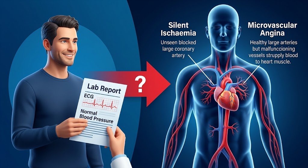
This article provides a comprehensive, evidence-based guide to coronary artery disease diagnosis, written for medical students, residents, healthcare professionals, and informed readers who want to understand how clinicians separate true CAD from the conditions that mimic it. If you want to understand why laboratory values can sometimes be misleading, our detailed guide to medical diagnosis explained in 15 powerful steps explains how clinicians interpret these results in context.
Key Diagnostic Facts About Coronary Artery Disease
The following table summarizes the most clinically important facts about coronary artery disease diagnosis. This is not a substitute for full clinical evaluation, but it provides a rapid reference for healthcare professionals and students.
| Diagnostic Feature | Key Fact |
|---|---|
| Disease | Coronary Artery Disease (CAD), also called Coronary Heart Disease (CHD) |
| Main clinical suspicion | Exertional chest discomfort or anginal equivalent relieved by rest, especially with cardiovascular risk factors |
| Most important symptoms | Substernal chest pressure, tightness, or heaviness provoked by exertion and relieved by rest; anginal equivalents include exertional dyspnoea, fatigue, or jaw/arm discomfort |
| Important risk factors | Age, male sex, smoking, diabetes, hypertension, dyslipidaemia, family history of premature CAD, obesity, sedentary lifestyle |
| First clinical step | Detailed history and physical examination, including assessment of chest pain typicality |
| Initial investigations | 12-lead resting ECG; blood tests including full blood count, fasting glucose or HbA1c, lipid profile, renal function, and high-sensitivity cardiac troponin (hs-cTn) |
| Important laboratory tests | hs-cTn (for acute coronary syndrome); lipid profile, HbA1c, and renal function for risk assessment |
| Important imaging | Coronary CT angiography (CCTA) for anatomical assessment; echocardiography for cardiac structure; stress imaging for functional assessment |
| Confirmatory test | Invasive coronary angiography (ICA) with fractional flow reserve (FFR) or instantaneous wave-free ratio (iFR) |
| Important differential diagnoses | Aortic stenosis, hypertrophic cardiomyopathy, oesophageal spasm, gastro-oesophageal reflux disease, musculoskeletal chest pain, anxiety, pulmonary embolism, pericarditis |
| Major red flags | Ongoing chest pain at rest, haemodynamic instability, ECG changes suggestive of acute ischaemia, troponin elevation, syncope, or new-onset heart failure symptoms |
| Specialist referral | Cardiology referral for patients with high clinical likelihood of obstructive CAD, abnormal non-invasive test results, or red-flag features |
| Diagnostic challenge | Up to 40% of patients with anginal symptoms have non-obstructive CAD (ANOCA/INOCA), requiring functional testing of the coronary microcirculation |
This table is a snapshot. Every parameter in it will be expanded in the dedicated sections below.
Step 1 — When Should Doctors Suspect Coronary Artery Disease?
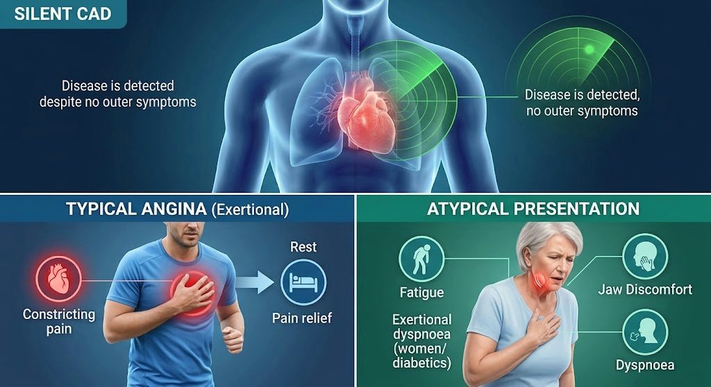
Typical Presentation: Coronary artery disease is often called the “silent killer” because most individuals with significant coronary atherosclerosis have no symptoms until plaque rupture or critical stenosis occurs. The condition is typically detected during evaluation of exertional chest discomfort, breathlessness, or an abnormal stress test. When symptoms do occur, they are usually predictable and reproducible. Typical angina is defined by three characteristic features: (1) substernal chest discomfort of a constricting, squeezing, or heavy quality; (2) provoked by physical exertion or emotional stress; and (3) relieved by rest or nitroglycerin within approximately five minutes.
Atypical Presentation: Older adults, women, and patients with diabetes may present with exertional dyspnoea, fatigue, nausea, or jaw or arm discomfort without classic chest pain. These anginal equivalents should be evaluated with the same diagnostic rigour as typical chest pain. Some patients present with symptoms of acute coronary syndrome, including rest pain, diaphoresis, or syncope.
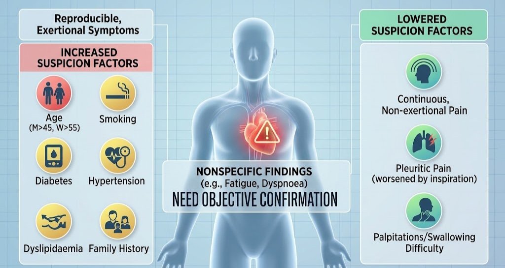
Risk Factors and Epidemiological Clues: Key risk factors that increase clinical suspicion include age (men >45 years, women >55 years), male sex, smoking, diabetes mellitus, hypertension, dyslipidaemia, family history of premature CAD (first-degree male relative <55 years, female relative <65 years), obesity, and sedentary lifestyle. These features increase the pretest likelihood of obstructive disease and guide the choice of diagnostic testing.
Features That Increase Clinical Suspicion: Clinical suspicion for CAD should be heightened when chest discomfort is exertional, reproducible, and relieved by rest. Features that lower suspicion include continuous or very prolonged pain, pain unrelated to activity, pleuritic pain worsened by inspiration, and pain associated with dizziness, palpitations, tingling, or difficulty swallowing.
Nonspecific Findings: Many findings associated with CAD are nonspecific, including fatigue, dyspnoea, and atypical chest discomfort. These symptoms occur commonly in non-cardiac conditions and should not be attributed to CAD without objective confirmation. If you want to understand how blood sugar clues can signal early metabolic disease — often coexisting with CAD — our detailed guide to diabetes blood sugar clues and early signs explains the diagnostic overlap in a clear, practical way.
Step 2 — Medical History
The medical history serves three purposes: to establish the diagnosis, to identify secondary causes, and to assess cardiovascular risk. Key areas to address include:
- Presenting complaint: Character, location, radiation, severity, duration, and frequency of symptoms.
- Onset and progression: When symptoms began, whether they are stable or progressive, and whether they occur at rest.
- Provoking and relieving factors: Exertion, emotional stress, rest, nitroglycerin, position, food, and breathing.
- Associated symptoms: Dyspnoea, diaphoresis, nausea, palpitations, syncope, or presyncope.
- Previous episodes: Prior chest pain, myocardial infarction, or revascularisation procedures.
- Previous diagnoses: Hypertension, diabetes, dyslipidaemia, peripheral artery disease, chronic kidney disease.
- Medications: Current and previous cardiovascular medications, including antiplatelets, statins, beta-blockers, and nitrates.
- Allergies: Particularly to contrast media, aspirin, or heparin.
- Family history: Premature CAD, sudden cardiac death, or inherited lipid disorders.
- Social history: Tobacco use, alcohol consumption, recreational drug use (especially cocaine), and occupational stress.
- Diet and physical activity: Dietary patterns, saturated fat intake, and exercise habits.
- Immunisation history: Influenza and pneumococcal vaccination status, relevant to overall cardiovascular risk management.
A structured approach to history-taking ensures that subtle clues are not missed and that the differential diagnosis remains broad until sufficient evidence accumulates. The 2025 ACC/AHA guideline emphasizes that when a new diagnosis of CAD is suspected, a comprehensive history and physical examination should be performed to inform management decisions.
For online earning ideas, freelancing, affiliate marketing, YouTube, Facebook monetization, and AI tips, visit ssthem.xyz for online earning and WhatsApp group links.
Physical Examination

Vital Signs and General Appearance: Blood pressure should be measured in both arms at the initial visit. A difference greater than 15 mmHg between arms warrants evaluation of the upper vasculature. Heart rate, respiratory rate, oxygen saturation, and body mass index (BMI) should be documented. Signs of distress, diaphoresis, pallor, or anxiety may be present in acute presentations.
Cardiovascular Examination: Auscultation for murmurs (aortic stenosis can mimic angina), carotid bruits, jugular venous pressure, peripheral pulses, and signs of heart failure such as raised JVP, basal crackles, or peripheral oedema. An S4 gallop may suggest left ventricular hypertrophy. A displaced apical impulse may indicate ventricular enlargement.
Specific Diagnostic Signs: Xanthelasma, tendon xanthomata, and corneal arcus suggest familial hypercholesterolaemia. Blood pressure differential between arms may suggest aortic dissection or subclavian stenosis. Diminished or delayed femoral pulses suggest coarctation of the aorta.
Findings Suggesting Alternative Diagnoses: Pleuritic rub (pericarditis), reproducible chest wall tenderness (musculoskeletal), epigastric tenderness (gastro-oesophageal reflux), or unilateral leg swelling (deep vein thrombosis with pulmonary embolism).
Findings suggesting complications include signs of heart failure (jugular venous distension, crackles, peripheral oedema), stroke (focal neurological deficits), or peripheral arterial disease (diminished pulses, trophic changes).
Red Flags That Should Not Be Ignored
The following table summarizes clinically meaningful warning signs that require urgent attention. These are not exaggerated risks but genuine indicators of potentially serious underlying pathology.
| Red Flag | Why It Matters | Possible Concern | Appropriate Action |
|---|---|---|---|
| Ongoing chest pain at rest >20 minutes | Suggests acute coronary syndrome | Unstable angina or myocardial infarction | Emergency department assessment; ECG within 10 minutes; hs-cTn |
| Haemodynamic instability (hypotension, tachycardia) | Indicates compromised cardiac output | Cardiogenic shock, massive MI | Emergency resuscitation; urgent cardiology |
| ST-segment elevation on ECG | Suggests acute coronary occlusion | STEMI | Immediate reperfusion therapy |
| New or worsening heart failure signs | May indicate ischaemic cardiomyopathy | Acute decompensated heart failure | Urgent echocardiography; cardiology referral |
| Syncope during exertion | Suggests severe outflow obstruction or arrhythmia | Critical aortic stenosis, severe CAD | Urgent cardiology assessment |
| Troponin elevation without obvious cause | Indicates myocardial injury | Myocarditis, pulmonary embolism, sepsis, or type 2 MI | Full diagnostic workup including imaging |
Differential Diagnosis
The following table outlines the most important conditions that can resemble coronary artery disease, along with their distinguishing features and key investigations.
| Differential Diagnosis | Similar Features | Distinguishing Features | Key Investigation |
|---|---|---|---|
| Aortic stenosis | Exertional chest pain, dyspnoea, syncope | Systolic murmur radiating to carotids; slow-rising pulse | Echocardiography |
| Hypertrophic cardiomyopathy | Exertional chest pain, dyspnoea, syncope | Systolic murmur worsening with Valsalva; family history | Echocardiography |
| Gastro-oesophageal reflux disease | Substernal burning, worse after meals and lying flat | Relieved by antacids; no exertional pattern | Trial of proton pump inhibitor; endoscopy if indicated |
| Musculoskeletal chest pain | Chest wall discomfort | Reproducible tenderness; worse with movement or palpation | Clinical examination; no specific test |
| Pulmonary embolism | Pleuritic chest pain, dyspnoea, tachycardia | Risk factors for VTE; sudden onset | D-dimer, CT pulmonary angiography |
| Pericarditis | Chest pain, worse with inspiration and lying flat | Pleuritic; relieved by sitting forward; pericardial rub | ECG (diffuse ST elevation), echocardiography |
| Anxiety/panic disorder | Chest tightness, palpitations, dyspnoea | Associated with psychological triggers; no exertional pattern | Clinical assessment; exclusion of organic causes |
Clinical reasoning behind these differences hinges on pattern recognition, risk factor profiling, and targeted investigation. A patient with exertional symptoms and a systolic murmur is more likely to have aortic stenosis than CAD. A patient with postprandial burning that worsens when lying down is more likely to have reflux. However, overlap exists, and multiple conditions can coexist.
Diagnostic Tests
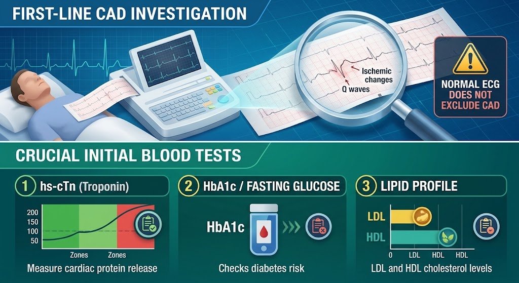
1. 12-Lead Resting Electrocardiogram (ECG): Ordered as the first-line test in any patient with suspected CAD. It assesses cardiac rhythm, identifies prior infarction (Q waves), and detects ischaemic changes (ST depression, T-wave inversion) that may be present even at rest. In acute presentations, a 12-lead ECG should be obtained within 10 minutes of first medical contact. Pathological Q waves, ST-segment depression, T-wave inversion in contiguous leads, or left bundle branch block may support the diagnosis. A completely normal ECG does not exclude CAD. Limitations include limited sensitivity for detecting CAD in stable patients; it cannot visualise coronary arteries. False positives and false negatives both occur. Abnormal ECG findings require further investigation with imaging or functional testing.
2. High-Sensitivity Cardiac Troponin (hs-cTn): Ordered to detect myocardial injury in acute presentations. hs-cTn assays detect smaller amounts of myocardial damage than conventional assays, enabling faster rule-in and rule-out of myocardial infarction. It measures cardiac troponin I or T, regulatory proteins released from damaged myocardium. A rise and/or fall in troponin above the 99th percentile upper reference limit, with at least one value above the limit, in the context of ischaemic symptoms or ECG changes, supports the diagnosis. A single normal troponin does not exclude ACS. Troponin elevation is not specific to CAD. Myocarditis, pulmonary embolism, sepsis, renal failure, and tachycardia can all cause elevation. Reference intervals vary between assays and laboratories.
3. Blood Tests Beyond Troponin: A standard laboratory panel in suspected CAD includes full blood count (anaemia can exacerbate angina and mimic CAD symptoms), fasting glucose or HbA1c (diabetes is a major risk factor and can be diagnosed concurrently), lipid profile (total cholesterol, LDL, HDL, and triglycerides), renal function (creatinine and estimated glomerular filtration rate, important before contrast imaging), liver function (baseline before statin initiation), thyroid function (hyperthyroidism can cause angina and tachycardia), and electrolytes (potassium and magnesium abnormalities can cause ECG changes). These tests do not diagnose CAD directly but inform risk assessment, identify exacerbating conditions, and guide safe investigation.
4. Echocardiogram for Coronary Artery Disease: Ordered to assess cardiac structure and function, identify regional wall motion abnormalities suggestive of prior infarction, and evaluate valve disease that may mimic or coexist with CAD. It measures chamber dimensions, wall thickness, ejection fraction, valvular function, and wall motion. Regional wall motion abnormalities in a coronary territory distribution may support the diagnosis. Normal wall motion and function do not exclude CAD, especially in stable disease without prior infarction. Resting echocardiography cannot detect coronary stenosis directly. It assesses the consequences of CAD, not the disease itself.
5. Exercise Stress Test for CAD: Ordered to provoke ischaemia through physical exertion and detect ECG changes, symptoms, or haemodynamic responses indicative of obstructive CAD. It measures ECG changes during graded exercise, exercise capacity, blood pressure response, and symptom reproduction. ST-segment depression ≥1 mm horizontal or downsloping, exercise-induced chest pain, or hypotension during exercise may support the diagnosis. Achievement of target heart rate without ECG changes or symptoms suggests a lower likelihood of obstructive CAD, but does not exclude it. Limitations include limited sensitivity and specificity compared with imaging stress tests. It cannot localise the ischaemic territory as precisely as imaging. Not suitable for patients with left bundle branch block, paced rhythm, or inability to exercise.
6. Stress Echocardiography: Ordered to combine exercise or pharmacological stress with echocardiographic imaging, detecting regional wall motion abnormalities that indicate ischaemia. It measures wall motion at rest and during stress. New or worsening regional wall motion abnormality during stress may support the diagnosis. Normal wall motion at rest and stress may argue against it. Limitations include operator dependence and image quality. Abnormal results generally prompt coronary angiography.
7. Coronary Artery Calcium Score: Ordered to quantify calcified plaque burden in asymptomatic individuals at intermediate risk, or in symptomatic patients when CAD is uncertain. It measures the Agatston score, derived from non-contrast CT. A score of 0 indicates very low 10-year event risk. A score >100 or >75th percentile reclassifies intermediate-risk individuals to high risk, benefiting from preventive therapy. Any score above 0 indicates coronary atherosclerosis, but not necessarily obstructive disease. Scores >400 indicate severe plaque burden. A calcium score of 0 strongly argues against significant calcified atherosclerosis but does not exclude non-calcified plaque. Limitations include inability to detect non-calcified plaque or stenosis.
8. Coronary CT Angiography (CCTA): Ordered as a first-line anatomical test in patients with low-to-intermediate pretest probability of obstructive CAD. It measures luminal stenosis, plaque burden, and plaque composition. CCTA has high sensitivity and negative predictive value for ruling out obstructive CAD. Stenosis ≥50% in a major epicardial vessel, or high-risk plaque features (positive remodelling, low-attenuation plaque, spotty calcification) may support the diagnosis. No stenosis or minimal plaque in a patient with low pretest probability may argue against it. Limitations include requirement for adequate heart rate control and renal function, contrast exposure, and less accuracy in heavily calcified vessels. Abnormal CCTA often leads to invasive angiography or functional testing.
9. Cardiac Stress Perfusion Imaging (MRI or PET): Ordered to assess myocardial perfusion and detect ischaemia with high spatial resolution. It measures myocardial blood flow at rest and stress. Reversible perfusion defect in a coronary territory may support the diagnosis. Normal perfusion at rest and stress may argue against it. Limitations include availability, cost, and contraindications (e.g., MRI in patients with certain implants).
10. Invasive Coronary Angiography (ICA): Ordered as the reference standard for anatomical assessment of coronary arteries and to guide revascularisation. It measures luminal diameter stenosis, plaque morphology, and coronary anatomy. Stenosis ≥70% (or ≥50% for left main) is considered obstructive. No stenosis or non-obstructive disease (stenosis <50%) may argue against it. Limitations include invasiveness, contrast exposure, small risk of complications, and visual estimation limitations. The angiogram is a “lumenogram” and does not visualise the vessel wall. How to interpret coronary angiography results requires integrating stenosis severity, plaque morphology, and functional assessment.
11. Fractional Flow Reserve (FFR) and Instantaneous Wave-Free Ratio (iFR): Ordered to assess the haemodynamic significance of intermediate coronary lesions (50–69% stenosis) during invasive angiography. FFR measures distal-to-proximal pressure ratio during maximal hyperaemia. iFR measures the ratio during the wave-free period of diastole. FFR ≤0.80 or iFR ≤0.89 indicates haemodynamically significant stenosis. FFR >0.80 or iFR >0.89 suggests the lesion is not flow-limiting. Limitations include requirement for adenosine or other hyperaemic agent. Not validated for all lesion types (e.g., left main, bifurcation).
12. Intravascular Imaging (IVUS and OCT): Ordered to characterise plaque morphology and optimise stent deployment during PCI. It measures plaque burden, calcification, dissection, and stent apposition. Useful in complex lesions, left main disease, or when angiography is ambiguous. Limitations include invasiveness, added procedural time, and cost. Not a first-line diagnostic test.
Laboratory Tests Table
| Test | Purpose | Possible Result | Interpretation | Limitations |
|---|---|---|---|---|
| hs-cTn | Detect myocardial injury | Rise/fall above 99th percentile | Supports MI in ischaemic context | Elevated in non-ischaemic conditions |
| Full blood count | Assess for anaemia | Anaemia may worsen angina | Baseline; not diagnostic of CAD | Nonspecific |
| Fasting glucose or HbA1c | Screen for diabetes | Elevated glucose or HbA1c indicates diabetes or prediabetes | Comorbidity affecting risk and treatment | HbA1c affected by hemoglobin variants |
| Lipid profile | Assess cardiovascular risk | Elevated LDL, low HDL increase risk | Guides statin therapy decisions | Fasting status may affect results |
| Serum creatinine with eGFR | Assess kidney function | Reduced eGFR indicates chronic kidney disease | Essential baseline; guides drug selection | eGFR equations vary by population |
| Thyroid-stimulating hormone | Screen for thyroid dysfunction | Hyperthyroidism or hypothyroidism may contribute to CAD | Treatable secondary cause | TSH may be normal in central thyroid disease |
| Electrolytes | Assess for abnormalities | Potassium and magnesium abnormalities can cause ECG changes | Guides safe investigation | Affected by medications, hydration |
| Liver function tests | Baseline before statin initiation | Abnormal results may affect drug selection | Guides safe therapy | Nonspecific |
Imaging Studies
Chest X-ray: Not used to diagnose CAD but may reveal cardiomegaly, pulmonary oedema, or alternative causes of chest pain. It provides a baseline assessment of cardiac silhouette and pulmonary vasculature.
Echocardiography: Assesses cardiac structure and function, identifying complications and alternative diagnoses. It is useful in patients with heart failure symptoms, murmurs, or abnormal ECG findings. Regional wall motion abnormalities in a coronary territory distribution may support the diagnosis. Normal wall motion and function do not exclude CAD.
Coronary CT Angiography: Provides detailed anatomical assessment and is a first-line test in low-to-intermediate pretest probability. It can visualise plaque burden and composition, which adds incremental value over stenosis alone. Plaque burden improves diagnostic accuracy; a percent atheroma volume threshold of 12.2% in intermediate stenosis achieves 88% diagnostic accuracy for ischaemic CAD.
Stress Imaging: Stress echo, perfusion MRI, or PET detects ischaemia and is useful when functional assessment is needed. PET has high diagnostic accuracy for ischaemic CAD. Each modality has strengths and limitations. CCTA is excellent for ruling out CAD but may overestimate stenosis in calcified vessels. Stress imaging assesses functional significance but may miss non-obstructive plaque.
Invasive Coronary Angiography: Remains the reference standard for anatomical assessment and guides revascularisation. It provides direct visualisation of the coronary lumen and allows measurement of FFR/iFR for functional assessment.
If you want to understand how hypertension diagnosis can overlap with CAD risk assessment, our detailed guide to hypertension diagnosis explained with BP readings and home monitoring explains the diagnostic criteria in a clear, practical way.
Specialized and Confirmatory Testing
Invasive coronary functional testing is recommended (Class 1) to confirm or rule out obstructive CAD or angina with non-obstructive coronary arteries (ANOCA). This includes assessment of coronary flow reserve, microvascular resistance, and acetylcholine provocative testing for vasomotor disorders.
Intravascular ultrasound (IVUS) and optical coherence tomography (OCT) characterise plaque and optimise PCI. They are used in complex lesions, left main disease, or when angiography is ambiguous.
Genetic testing is not routine in CAD but may be considered in familial hypercholesterolaemia or premature CAD. Biomarkers beyond troponin, such as B-type natriuretic peptide (BNP), have prognostic value but are not diagnostic.
Diagnostic Criteria
There is no single universally accepted diagnostic criterion for CAD. Diagnosis integrates clinical presentation, test results, and anatomical or functional evidence of obstructive disease. Obstructive CAD is generally defined as stenosis ≥70% in a major epicardial vessel or ≥50% in the left main. Non-obstructive CAD includes stenosis <50% or ANOCA.
Acute coronary syndrome is diagnosed when there is evidence of myocardial ischaemia with troponin rise/fall (NSTEMI), ST elevation (STEMI), or unstable angina (ischaemic symptoms without troponin elevation). The 2025 ACC/AHA guideline provides updated diagnostic distinctions using ECG, troponin, and imaging.
Pretest probability assessment is integral. The 2024 ESC guideline recommends a risk factor-weighted clinical likelihood model to categorise patients as very low (≤5%), low-to-moderate (>5–50%), or high (>50–85%). CCTA is recommended for low-to-moderate likelihood; functional imaging or invasive testing for high likelihood.
Diagnostic Algorithm
| Step | Action | Decision Point |
|---|---|---|
| 1. Symptoms | Assess chest pain typicality and anginal equivalents | Typical, atypical, or non-anginal |
| 2. Clinical suspicion | Identify risk factors and red flags | Low, intermediate, or high suspicion |
| 3. History | Detailed symptom characterisation | Exertional pattern, relief with rest |
| 4. Physical examination | Vital signs, murmurs, heart failure signs | Alternative diagnoses, complications |
| 5. Initial tests | ECG, hs-cTn, blood tests | Acute vs. stable presentation |
| 6. Interpretation | Integrate findings | Pretest probability |
| 7. Differential diagnosis | Exclude mimics | Aortic stenosis, reflux, PE |
| 8. Confirmatory testing | CCTA, stress imaging, or ICA | Anatomical vs. functional |
| 9. Severity/staging | Stenosis severity, plaque burden, ischaemic burden | Obstructive vs. non-obstructive |
| 10. Specialist evaluation | Cardiology referral | Revascularisation vs. medical therapy |
| 11. Follow-up | Risk factor management, symptom monitoring | Ongoing assessment |
Severity and Staging
CAD severity is classified anatomically and functionally. Mild: Non-obstructive plaque (<50% stenosis). Managed with risk factor modification. Moderate: Intermediate stenosis (50–69%). Functional assessment (FFR/iFR) determines significance. Severe: Obstructive stenosis (≥70%, or ≥50% left main). Revascularisation generally considered.
High-risk features include left main disease, proximal LAD disease, three-vessel disease, and high plaque burden on CCTA. These influence prognosis and revascularisation decisions.
Special Populations
Older adults: May present with atypical symptoms, including dyspnoea, confusion, or falls. Diagnostic thresholds for troponin may differ. Comorbidities increase procedural risk.
Women: More likely to present with atypical symptoms. Non-obstructive CAD and ANOCA are more common. Diagnostic imaging may be less accurate in smaller vessels.
Patients with diabetes: Often have silent ischaemia and diffuse multi-vessel disease. Screening for CAD in asymptomatic diabetes remains controversial.
Patients with chronic kidney disease: Contrast exposure requires careful risk-benefit assessment. FFR/iFR can be performed with minimal contrast.
Pregnancy: Exercise stress testing is preferred over pharmacological stress. Radiation exposure should be minimised. CCTA is generally avoided.
False Positive and False Negative Results
False Positive: A false-positive result occurs when a test suggests CAD when the condition is actually absent. The most common example is exercise ECG, where ST changes occur without obstructive CAD. This can result from left ventricular hypertrophy, electrolyte abnormalities, or digoxin use. CCTA can overestimate stenosis in heavily calcified vessels. Troponin elevation from non-ischaemic causes can also produce false-positive results. Overdiagnosis can lead to unnecessary invasive procedures and associated adverse effects.
False Negative: A false-negative result occurs when a test suggests normal coronary arteries when CAD is present. A single normal troponin can be normal in early MI. Exercise ECG can be normal in single-vessel disease. CCTA can miss non-calcified plaque if image quality is poor. Underdiagnosis leaves patients at risk for preventable complications.
Clinicians use pretest probability to interpret results. A positive test in a high-probability patient is more likely to be a true positive. A negative test in a low-probability patient is more likely to be a true negative. Confirmatory testing is used when results are discordant with clinical suspicion.
The most useful test is not necessarily the most advanced test. Sometimes the most important diagnostic tool is a carefully taken history and a properly interpreted ECG.
Common Diagnostic Mistakes in Coronary Artery Disease
- Anchoring bias: Fixating on an initial diagnosis (e.g., musculoskeletal pain) and ignoring evolving evidence.
- Premature diagnostic closure: Stopping the diagnostic process once one abnormality is found, without excluding alternatives.
- Ignoring red flags: Dismissing ongoing chest pain at rest or haemodynamic instability.
- Over-relying on one laboratory test: Treating a normal troponin as definitive exclusion of ACS.
- Misunderstanding reference ranges: Interpreting troponin or lipid values without considering assay-specific intervals.
- Ignoring pretest probability: Ordering advanced tests indiscriminately, leading to false positives and unnecessary procedures.
- Failing to consider differential diagnoses: Attributing all chest pain to CAD without excluding aortic stenosis, reflux, or pulmonary embolism.
- Inappropriate test selection: Using exercise ECG in patients with left bundle branch block, where it is uninterpretable.
- Misinterpreting false-positive results: Performing invasive angiography based on a false-positive non-invasive test.
- Failing to reassess an evolving patient: Not repeating ECG or troponin when symptoms persist or recur.
Hypothetical Clinical Cases
These are hypothetical educational cases and do not represent real patients.
Case 1: Exertional Chest Pain in a 58-Year-Old Man
Presentation: A 58-year-old man with a 20-pack-year smoking history, hypertension, and dyslipidaemia presents with three months of substernal chest pressure occurring during brisk walking and relieved within five minutes of rest.
Initial Differential: Stable angina, gastro-oesophageal reflux, musculoskeletal pain.
History: Symptoms are consistently provoked by exertion and relieved by rest. No rest pain, no dyspnoea at rest. Father had myocardial infarction at age 60.
Examination: Blood pressure 148/92 mmHg, heart rate 78 bpm. No murmurs. Peripheral pulses intact.
Investigations: Resting ECG normal. Blood tests: LDL 4.2 mmol/L, HbA1c 5.9%, creatinine normal.
Test Results: Exercise stress test shows 1.5 mm ST depression in leads V4–V6 at 7 METs, associated with chest pain.
Clinical Reasoning: Typical angina with intermediate pretest probability. Exercise ECG positive for ischaemia.
Diagnosis: Stable angina due to obstructive CAD.
Key Diagnostic Lesson: Exercise stress testing remains a valuable first-line functional test in patients who can exercise and have interpretable ECGs.
Case 2: Exertional Dyspnoea in a 62-Year-Old Woman with Diabetes
Presentation: A 62-year-old woman with type 2 diabetes and hypertension presents with six weeks of exertional dyspnoea and fatigue, without chest pain.
Initial Differential: CAD with anginal equivalent, heart failure, anaemia.
History: Symptoms occur with moderate exertion and resolve with rest. No orthopnoea or paroxysmal nocturnal dyspnoea.
Examination: BMI 32, blood pressure 152/88 mmHg. No murmurs, no crackles.
Investigations: ECG shows T-wave inversion in leads V4–V6. hs-cTn normal. HbA1c 8.1%. Echocardiogram shows mild left ventricular hypertrophy with preserved ejection fraction.
Test Results: CCTA shows 60% stenosis in the proximal LAD with high-risk plaque features.
Clinical Reasoning: Anginal equivalent in a patient with diabetes. CCTA identifies obstructive CAD with high-risk plaque.
Diagnosis: CAD with anginal equivalent presentation.
Key Diagnostic Lesson: Exertional dyspnoea in patients with diabetes should be evaluated as a potential anginal equivalent.
Case 3: Acute Chest Pain in a 45-Year-Old Man with Normal Initial ECG
Presentation: A 45-year-old man presents to the emergency department with one hour of central chest pressure that started at rest.
Initial Differential: Acute coronary syndrome, pulmonary embolism, pericarditis.
History: Sudden onset at rest, associated with diaphoresis. No prior cardiac history.
Examination: Heart rate 95 bpm, blood pressure 135/85 mmHg, diaphoretic.
Investigations: Initial ECG normal. Initial hs-cTn 8 ng/L (below 99th percentile). Repeat hs-cTn at 1 hour: 45 ng/L (significant rise).
Test Results: Invasive coronary angiography shows 95% stenosis in the right coronary artery.
Clinical Reasoning: Serial troponin sampling is essential. A single normal troponin does not exclude ACS.
Diagnosis: Non-ST elevation myocardial infarction (NSTEMI).
Key Diagnostic Lesson: Serial hs-cTn testing with a 1-hour algorithm enables rapid and accurate rule-in/rule-out of MI.
Patient-Friendly Diagnosis Guide
What Should a Patient Expect During Diagnosis?
If your doctor suspects coronary artery disease, the diagnostic process typically unfolds in several steps:
Consultation and history: Your doctor will ask detailed questions about your symptoms: when they occur, what provokes them, what relieves them, and how long they last. Be specific and honest about your symptoms, medications, and lifestyle.
Physical examination: Your doctor will check your blood pressure, heart rate, and listen to your heart and lungs. They may also check pulses in your legs and neck.
Blood tests: You will likely have blood drawn to check for anaemia, diabetes, cholesterol levels, kidney function, and, if you are having acute symptoms, cardiac troponin. These tests help identify risk factors and exclude other causes.
ECG: A simple, painless test that records your heart’s electrical activity. It takes less than five minutes.
Imaging: Depending on your risk and symptoms, your doctor may order a coronary CT angiography (a CT scan of the heart arteries), a stress test (exercise or pharmacological), or an echocardiogram. These tests assess the structure and function of your heart and arteries.
Invasive tests: If non-invasive tests suggest significant disease, your doctor may recommend coronary angiography. This involves threading a thin tube through a blood vessel to the heart to inject contrast dye and visualise the arteries. It is performed under local anaesthesia.
Waiting for results: Some tests take time to analyse. Your doctor will explain when to expect results and what they mean.
Follow-up: Based on your results, your doctor will discuss whether you need treatment, further tests, or lifestyle changes. Diagnosis is often a process, not a single test.
When to Seek Urgent Medical Care
Seek emergency medical care immediately if you experience:
- Chest pain or pressure that lasts more than a few minutes, especially at rest
- Chest pain with sweating, nausea, or shortness of breath
- Fainting or severe dizziness
- Rapid or irregular heartbeat with chest discomfort
- Sudden severe shortness of breath
Do not drive yourself to the hospital. Call emergency services. Do not attempt to self-diagnose or self-treat. This article is for education and does not replace professional medical assessment.
1. A 58-year-old man presents with substernal chest pressure provoked by exertion and relieved by rest within 5 minutes. His resting ECG is normal. What is the most appropriate first-line diagnostic test?
A. Coronary CT angiography
B. Exercise stress test
C. Invasive coronary angiography
D. Coronary artery calcium score
Correct Answer: B
Explanation: Exercise stress testing is the appropriate first-line functional test in patients with intermediate pretest probability who can exercise and have an interpretable ECG.
2. A 45-year-old woman presents to the emergency department with 2 hours of chest pain. Her initial hs-cTn is 5 ng/L (normal). What is the next step?
A. Reassure and discharge
B. Repeat hs-cTn in 1 hour
C. Order coronary CT angiography
D. Start thrombolysis
Correct Answer: B
Explanation: Serial hs-cTn sampling is essential. A single normal troponin does not exclude ACS. A 1-hour repeat is recommended with high-sensitivity assays.
3. Which ECG finding is most suggestive of prior myocardial infarction?
A. ST elevation
B. T-wave inversion
C. Pathological Q waves
D. Left bundle branch block
Correct Answer: C
Explanation: Pathological Q waves indicate myocardial necrosis and are a marker of prior infarction.
4. A 62-year-old man with a calcium score of 450 asks what this means. What is the most accurate response?
A. He has obstructive CAD
B. He has severe calcified plaque burden and high cardiovascular risk
C. He needs immediate angiography
D. He has no significant disease
Correct Answer: B
Explanation: A calcium score >400 indicates severe calcified plaque burden and high risk. It does not diagnose obstructive CAD but reclassifies risk.
5. Which test is the reference standard for anatomical assessment of coronary stenosis?
A. Coronary CT angiography
B. Stress echocardiography
C. Invasive coronary angiography
D. Coronary artery calcium score
Correct Answer: C
Explanation: Invasive coronary angiography is the reference standard for anatomical assessment of coronary arteries.
6. A patient has a 60% stenosis in the LAD on angiography. What is the next most appropriate step?
A. Immediate stenting
B. Fractional flow reserve measurement
C. Medical therapy alone
D. Repeat angiography in 6 months
Correct Answer: B
Explanation: Intermediate lesions (50–69%) require functional assessment with FFR or iFR to determine haemodynamic significance.
7. An FFR value of 0.75 indicates:
A. Non-significant stenosis
B. Haemodynamically significant stenosis
C. Normal coronary flow
D. Need for repeat measurement
Correct Answer: B
Explanation: FFR ≤0.80 indicates haemodynamically significant stenosis.
8. A 70-year-old woman presents with exertional dyspnoea and fatigue but no chest pain. What is this presentation called?
A. Typical angina
B. Atypical angina
C. Anginal equivalent
D. Non-anginal chest pain
Correct Answer: C
Explanation: Exertional dyspnoea, fatigue, or weakness without chest pain may be an anginal equivalent, especially in older adults and women.
9. Which of the following is a major risk factor for CAD?
A. Hypothyroidism
B. Diabetes mellitus
C. Asthma
D. Osteoporosis
Correct Answer: B
Explanation: Diabetes mellitus is a major modifiable risk factor for CAD.
10. What is the recommended time for obtaining a 12-lead ECG in suspected ACS?
A. Within 10 minutes of first medical contact
B. Within 30 minutes
C. Within 1 hour
D. Within 2 hours
Correct Answer: A
Explanation: The 2025 ACC/AHA guideline recommends a 12-lead ECG within 10 minutes of first medical contact.
11. A 55-year-old man has a normal resting ECG but typical angina. What does this mean?
A. CAD is excluded
B. Further testing is needed
C. He has non-cardiac chest pain
D. He needs immediate angiography
Correct Answer: B
Explanation: A normal resting ECG does not exclude CAD. Further testing with functional or anatomical imaging is needed.
12. Which of the following is a limitation of coronary CT angiography?
A. Cannot visualise coronary arteries
B. Overestimates stenosis in heavily calcified vessels
C. Requires general anaesthesia
D. Has low negative predictive value
Correct Answer: B
Explanation: CCTA can overestimate stenosis in heavily calcified vessels, reducing specificity.
13. A patient with suspected ACS has a troponin level above the 99th percentile with a rise and fall. What does this indicate?
A. Myocardial injury
B. Normal finding
C. Pulmonary embolism
D. Stable angina
Correct Answer: A
Explanation: A rise and fall in troponin above the 99th percentile indicates myocardial injury, which may be ischaemic (MI) or non-ischaemic.
14. Which test assesses the functional significance of a coronary stenosis during angiography?
A. Coronary calcium score
B. Fractional flow reserve
C. Resting ECG
D. Chest X-ray
Correct Answer: B
Explanation: FFR measures the pressure ratio across a stenosis during hyperaemia to assess functional significance.
15. A 68-year-old man with diabetes presents with exertional dyspnoea. What is the most appropriate initial diagnostic approach?
A. Reassure as non-cardiac
B. Evaluate as possible anginal equivalent
C. Order chest X-ray only
D. Start antibiotics
Correct Answer: B
Explanation: Exertional dyspnoea in a patient with diabetes should be evaluated as a potential anginal equivalent.
16. Which imaging modality provides both anatomical and functional assessment of CAD?
A. Chest X-ray
B. Coronary CT angiography with CT-FFR
C. Resting echocardiography
D. Abdominal ultrasound
Correct Answer: B
Explanation: CCTA provides anatomical assessment; CT-FFR adds functional information about lesion significance.
17. What is the pretest probability category for a 50-year-old man with typical angina and no risk factors?
A. Very low (≤5%)
B. Low-to-moderate (5–50%)
C. High (>50–85%)
D. Very high (>85%)
Correct Answer: B
Explanation: A 50-year-old man with typical angina but no risk factors falls into the low-to-moderate likelihood category, where CCTA is recommended.
18. Which of the following is a false positive cause of exercise ECG?
A. Single-vessel disease
B. Left ventricular hypertrophy
C. Submaximal exercise
D. Beta-blocker use
Correct Answer: B
Explanation: Left ventricular hypertrophy can cause ST-T changes that mimic ischaemia, leading to false-positive exercise ECG results.
19. A patient has a calcium score of 0. What does this indicate?
A. No atherosclerosis at all
B. Very low 10-year event risk
C. Obstructive CAD
D. Need for immediate angiography
Correct Answer: B
Explanation: A calcium score of 0 indicates very low 10-year event risk but does not exclude non-calcified plaque.
20. Which of the following is a red flag in a patient with chest pain?
A. Pain reproduced by palpation
B. Pain relieved by antacids
C. Ongoing chest pain at rest
D. Pain lasting seconds
Correct Answer: C
Explanation: Ongoing chest pain at rest suggests acute coronary syndrome and requires urgent assessment.
21. What is the primary role of echocardiography in CAD diagnosis?
A. Directly visualise coronary stenosis
B. Assess cardiac structure and function
C. Measure coronary flow reserve
D. Detect coronary calcification
Correct Answer: B
Explanation: Echocardiography assesses cardiac structure, function, and wall motion, which may be abnormal in CAD.
22. A 72-year-old woman with atypical chest pain and a normal exercise ECG. What is the next best step?
A. Reassure and discharge
B. Consider CCTA or stress imaging
C. Immediate angiography
D. Start thrombolysis
Correct Answer: B
Explanation: In patients with atypical symptoms and a normal exercise ECG but ongoing clinical suspicion, CCTA or stress imaging may provide additional diagnostic information.
23. Which of the following is a limitation of hs-cTn testing?
A. Cannot detect myocardial injury
B. Elevated in non-ischaemic conditions
C. Requires fasting
D. Only available in tertiary centres
Correct Answer: B
Explanation: hs-cTn is elevated in myocarditis, pulmonary embolism, sepsis, and renal failure, reducing specificity for CAD.
24. What is the diagnostic threshold for FFR indicating significant stenosis?
A. ≤0.50
B. ≤0.60
C. ≤0.80
D. ≤0.90
Correct Answer: C
Explanation: FFR ≤0.80 indicates haemodynamically significant stenosis.
25. A patient with ANOCA (angina with non-obstructive coronary arteries) has normal angiography. What is the next appropriate diagnostic step?
A. Reassure and discharge
B. Invasive coronary functional testing
C. Start thrombolysis
D. Repeat angiography in 1 year
Correct Answer: B
Explanation: Invasive coronary functional testing (coronary flow reserve, microvascular resistance, acetylcholine testing) is recommended to identify coronary microvascular dysfunction or vasomotor disorders in ANOCA.
Question. How is coronary artery disease diagnosed?
Answer : CAD is diagnosed through a combination of clinical assessment, ECG, blood tests, and imaging or functional tests. The specific tests depend on whether the presentation is acute or stable, and on the pretest probability of obstructive disease.
Question. What is the first test for coronary artery disease?
Answer : The first test is usually a 12-lead resting ECG, along with a detailed history and physical examination. Blood tests including hs-cTn are added in acute presentations.
Question. What blood tests are used for CAD?
Answer : hs-cTn detects myocardial injury. Lipid profile, HbA1c, full blood count, renal function, and thyroid function assess risk factors and exclude exacerbating conditions.
Question. Can CAD be diagnosed without a specific test?
Answer : CAD cannot be reliably diagnosed on symptoms alone. Clinical assessment guides the choice of tests, but objective evidence from ECG, imaging, or functional testing is required.
Question. What does a positive test mean?
Answer : A positive test suggests CAD but does not confirm it in isolation. Further testing is often needed to confirm obstructive disease and assess severity.
Question. Can diagnostic tests be falsely negative?
Answer : Yes. A normal resting ECG, a single normal troponin, or a normal exercise ECG do not exclude CAD.
Question. Can diagnostic tests be falsely positive?
Answer : Yes. ST changes on exercise ECG, troponin elevation from non-ischaemic causes, and CCTA overestimation of calcified stenosis can all produce false-positive results.
Question. When is imaging needed?
Answer : Imaging is needed when non-invasive assessment suggests CAD or when symptoms persist despite normal initial tests. CCTA is first-line for low-to-intermediate pretest probability. Stress imaging is used when functional assessment is needed.
Question. When is invasive angiography needed?
Answer : Invasive angiography is needed for high pretest probability, abnormal non-invasive tests, acute coronary syndrome, or when revascularisation is being considered.
Question. When should a specialist be consulted?
Answer : Cardiology referral is appropriate for patients with high clinical likelihood of obstructive CAD, abnormal non-invasive test results, red-flag features, or when invasive testing is being considered.
Question. When should urgent medical care be sought?
Answer : Urgent care is needed for ongoing chest pain at rest, chest pain with sweating or breathlessness, fainting, or severe dizziness.
Question. Can an ECG detect coronary artery disease?
Answer : An ECG can detect signs of prior infarction or ischaemia but cannot visualise coronary arteries. A normal ECG does not exclude CAD.
Question. What is the best test for coronary artery disease?
Answer : There is no single best test. The optimal test depends on pretest probability, patient characteristics, and whether anatomical or functional assessment is needed.
Question. How is coronary artery blockage diagnosed?
Answer : Coronary artery blockage is diagnosed by anatomical imaging showing stenosis, confirmed as functionally significant by FFR/iFR or stress testing.
Question. How do doctors diagnose CAD early?
Answer : Early diagnosis relies on recognising symptoms including anginal equivalents, assessing risk factors, and using appropriate non-invasive testing such as CCTA in patients with low-to-intermediate pretest probability.
Latest Evidence & References
- Roth GA, Mensah GA, Johnson CO, et al. Global Burden of Cardiovascular Diseases and Risk Factors, 1990–2019: Update From the GBD 2019 Study. Journal of the American College of Cardiology. 2020;76(25):2982–3021.
- National Institute for Health and Care Excellence (NICE). Chest pain of recent onset: assessment and diagnosis. Clinical guideline CG95. Updated 2016.
- RACGP. Chest pain. Australian Journal of General Practice. 2024;53(7):437–442.
- Gulati M, Levy PD, Mukherjee D, et al. 2021 AHA/ACC/ASE/CHEST/SAEM/SCCT/SCMR Guideline for the Evaluation and Diagnosis of Chest Pain. Circulation. 2021;144(22):e368–e454.
- Arnett DK, Blumenthal RS, Albert MA, et al. 2019 ACC/AHA Guideline on the Primary Prevention of Cardiovascular Disease. Circulation. 2019;140(11):e596–e646.
- Rao SV, O’Donoghue ML, Ruel M, et al. 2025 ACC/AHA/ACEP/NAEMSP/SCAI Guideline for the Management of Patients With Acute Coronary Syndromes. Circulation. 2025.
- McDermott M, et al. Cardiac troponin at the point of care in acute and chronic coronary syndromes. Diabetes, Obesity and Metabolism. 2025;27(Suppl 8):47–58.
- Pinto-Sietsma SJ, Velthuis BK, Nurmohamed NS, et al. Computed tomography and coronary artery calcium score for screening of coronary artery disease. Netherlands Heart Journal. 2024;32(11):371–377.
- Kero T, et al. Plaque burden improves the detection of ischemic CAD over stenosis from coronary computed tomography angiography. International Journal of Cardiovascular Imaging. 2025;41(6):1131–1140.
- Patel MR, Calhoon JH, Dehmer GJ, et al. ACC/AATS/AHA/ASE/ASNC/SCAI/SCCT/STS 2017 Appropriate Use Criteria for Coronary Revascularization in Patients With Stable Ischemic Heart Disease. Journal of the American College of Cardiology. 2017;69(17):2212–2241.
- Davies JE, Sen S, Dehbi HM, et al. Use of the Instantaneous Wave-free Ratio or Fractional Flow Reserve in PCI. New England Journal of Medicine. 2017;376(19):1824–1834.
- Gurgoglione FL, Niccoli G. What is New from the 2024 European Society of Cardiology Congress on the Management of Chronic Coronary Syndromes? European Cardiology Review. 2024;19:e23.
- Knuuti J, Wijns W, Saraste A, et al. 2019 ESC Guidelines for the diagnosis and management of chronic coronary syndromes. European Heart Journal. 2020;41(3):407–477.
- American College of Cardiology. Top Takeaways From the 2025 ACC/AHA Acute Coronary Syndrome Guideline. 2025.
Medical Information Disclaimer: The information provided in this article is for educational and informational purposes only and is intended for healthcare professionals, medical students, and informed general readers. It does not constitute medical advice, diagnosis, or treatment recommendations. Coronary artery disease is a medical condition that should be evaluated and managed by a qualified healthcare provider. Treatment decisions, including medication selection, revascularisation, and monitoring, depend on the patient’s individual circumstances, comorbidities, and clinical judgment. Readers should not use this information to self-diagnose, self-treat, or make clinical decisions without appropriate professional consultation. If you have concerns about your heart health, seek evaluation from a qualified healthcare professional. The authors and publishers of this article do not assume any liability for any adverse effects or consequences resulting from the use or misuse of the information provided herein.
discover well-organized, informative content. For health and beauty tips, visit ssthem.net for health and beauty tips.



2 Comments