TB or Not TB? 3 Life-Saving Steps to a Fast, Accurate Diagnosis
TB or Not TB: 3 Game-Changing Steps to a Fast Tuberculosis Diagnosis
A 28-year-old healthcare worker presents with a three-week history of productive cough, low-grade fever, and drenching night sweats. She has lost four kilograms. The first sputum test is reported as “smear-negative.” Her chest X-ray shows a subtle right upper lobe opacity. The initial interferon-gamma release assay is positive. It would be easy to conclude that this is latent tuberculosis infection and simply reassure her. Yet the real diagnostic question has only just begun: what evidence can separate active pulmonary tuberculosis from latent infection, non-tuberculous mycobacterial disease, fungal infection, sarcoidosis, or a dozen other conditions that can mimic upper lobe lung disease? The answer lies not in a single test, but in a disciplined diagnostic process that integrates history, examination, laboratory data, imaging, and careful clinical reasoning.
This is a hypothetical educational case, but it reflects a daily reality in clinical practice. Tuberculosis (TB) remains one of the world’s leading infectious causes of death, yet its diagnosis is rarely as simple as a single abnormal chest X-ray. The consequences of both overdiagnosis and underdiagnosis are substantial: unnecessary treatment and drug toxicity on one hand, and preventable transmission, respiratory failure, and death on the other. Accurate diagnosis requires a systematic approach that integrates clinical history, physical examination, laboratory testing, imaging, and, when appropriate, microbiological confirmation.
A normal chest X-ray does not always mean the disease is absent. A negative sputum test does not always mean the patient is not infectious. Diagnosis is a process, not a single number on a laboratory report.
This article provides a comprehensive, evidence-based guide to Tuberculosis Diagnosis, written for medical students, residents, healthcare professionals, and informed readers who want to understand how clinicians separate true TB from the conditions that mimic it. If you want to understand why laboratory values and imaging findings can sometimes be misleading, our detailed guide to medical diagnosis explained in 15 powerful steps explains how clinicians interpret these results in context.
Key Diagnostic Facts About Tuberculosis
The following table summarizes the most clinically important facts about tuberculosis diagnosis. This is not a substitute for full clinical evaluation, but it provides a rapid reference for healthcare professionals and students.
| Diagnostic Feature | Key Fact |
|---|---|
| Disease | Tuberculosis (TB), caused by Mycobacterium tuberculosis complex; pulmonary and extrapulmonary forms exist |
| Main clinical suspicion | Persistent cough >2 weeks, fever, night sweats, unintentional weight loss, hemoptysis; compatible epidemiological risk factors |
| Most important symptoms | Productive cough lasting more than two weeks, hemoptysis, fever, drenching night sweats, weight loss, fatigue, anorexia |
| Important risk factors | Close contact with infectious TB, residence in or travel to high-burden regions, HIV infection, immunosuppressive therapy (especially TNF-α inhibitors), diabetes, silicosis, end-stage renal disease, imprisonment, homelessness |
| First clinical step | Detailed history including symptom duration, exposure risk, medical history, and medication review |
| Initial investigations | Chest radiograph, sputum collection for molecular testing (Xpert MTB/RIF Ultra or Truenat), AFB smear, mycobacterial culture |
| Important laboratory tests | Molecular WHO-recommended rapid diagnostics (mWRDs), AFB smear microscopy, liquid culture (MGIT), line probe assay for drug resistance, urine lipoarabinomannan (LAM) in PLHIV, C-reactive protein (CRP) for screening |
| Important imaging | Posterior-anterior chest radiograph is the standard initial imaging modality; CT for equivocal or complicated cases; MRI for spinal and musculoskeletal TB |
| Confirmatory test | Positive culture or positive nucleic acid amplification test (NAAT) from a clinical specimen |
| Important differential diagnoses | Community-acquired pneumonia, lung malignancy, non-tuberculous mycobacterial infection, sarcoidosis, fungal infection, bronchiectasis, pleural mesothelioma |
| Major red flags | Massive hemoptysis, severe breathlessness, confusion or altered consciousness, neck stiffness with headache, miliary pattern on chest X-ray, rapid clinical deterioration |
| Specialist referral | Infectious disease, respiratory medicine, or TB specialist for drug-resistant or complicated cases, extrapulmonary TB, pediatric TB, TB in pregnancy, or TB in immunocompromised patients |
| Diagnostic challenge | Paucibacillary disease, immunocompromised states, extrapulmonary TB, and distinguishing latent from active infection |
This table is a snapshot. Every parameter in it will be expanded in the dedicated sections below.
Step 1 — When Should Doctors Suspect Tuberculosis?
Typical Presentation: 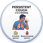 Tuberculosis should enter the differential diagnosis when a patient presents with a compatible clinical syndrome and has epidemiological risk factors that increase the pretest probability of infection. The classic triad includes a persistent cough lasting more than two to three weeks, fever, and unintentional weight loss. Cough may be dry initially but often becomes productive with purulent or blood-streaked sputum. Hemoptysis (coughing up blood) is a concerning feature that warrants prompt evaluation. Night sweats — drenching sweats that soak bedclothes — are highly suggestive when present alongside other symptoms.
Early Symptoms: Early TB can be insidious. Fatigue, malaise, low-grade fever, and a mild cough may be the only presenting features. This is why TB is sometimes called “the great imitator” — its early symptoms overlap with many common conditions. A cough lasting more than two weeks that does not respond to conventional antibiotics should prompt consideration of TB.
Characteristic Features That Increase Clinical Suspicion: These include upper lobe consolidation or cavitation on chest imaging, focal crackles or bronchial breathing on auscultation, new hypoxaemia, and a compatible systemic illness. However, none of these findings is perfectly sensitive or specific. The absence of crackles does not exclude TB. A normal chest X-ray in the early stages does not exclude TB, particularly in immunocompromised patients.
Atypical Presentation: Atypical presentations are particularly common in immunocompromised patients, older adults, and children. In people living with HIV (PLHIV), especially those with CD4 counts below 200 cells/mm³, TB may present with fewer classic respiratory symptoms and more systemic or disseminated features. Older adults may present with unexplained weight loss, functional decline, or anorexia without fever or cough. In children, TB may present with failure to thrive, persistent cough, and lymphadenopathy.
Duration and Course: TB symptoms typically evolve over weeks to months. A more acute course over hours to days should raise suspicion for community-acquired pneumonia or pulmonary embolism. Aspiration pneumonia may follow a witnessed aspiration event or occur in patients with dysphagia, altered consciousness, or neurological disease.
Epidemiological Clues: Close contact with a person known to have infectious TB, residence in or travel to a high-burden country, HIV infection, immunosuppressive therapy, diabetes mellitus, silicosis, end-stage renal disease on dialysis, history of imprisonment, homelessness, or residence in long-term care facilities all increase the pretest probability of TB. These clues do not replace systematic evaluation, but they can meaningfully alter the diagnostic approach.
Nonspecific Findings: Fatigue, myalgia, headache, and general malaise are common in both TB and non-TB conditions. If you want to understand how another major risk factor — diabetes — can quietly damage blood vessels and impair immune defence for years before producing symptoms, our detailed guide to diabetes blood sugar clues and early signs explains the diagnostic overlap in a clear, practical way.
Step 2 — Medical History
The medical history is the foundation of TB diagnosis. The information gathered during history-taking determines which tests are ordered, how results are interpreted, and what alternative diagnoses are considered. A focused but thorough history should elicit the following:
- Presenting complaint: What exactly happened? The patient’s own words should be recorded, followed by a structured characterisation of each symptom.
- Onset and duration: Acute onset over hours suggests bacterial aetiology or pulmonary embolism. Subacute onset over days is typical of viral pneumonia. A more chronic course over weeks suggests tuberculosis or malignancy.
- Progression and severity: Is the cough worsening? Is there hemoptysis? Are there night sweats? Is there weight loss? These simple questions provide rapid assessment of disease activity.
- Associated symptoms: Sputum colour and volume, haemoptysis, night sweats, weight loss, pleuritic pain, and extrapulmonary symptoms such as lymphadenopathy or joint pain should be documented.
- Previous episodes and diagnoses: Prior TB infection or disease, previous TB treatment and adherence, previous positive tuberculin skin test (TST) or interferon-gamma release assay (IGRA), and previous chest X-ray findings are all relevant.
- Medications and allergies: Immunosuppressive medications, particularly tumor necrosis factor-alpha (TNF-α) inhibitors (infliximab, adalimumab, etanercept), corticosteroids, chemotherapy agents, and any prior anti-TB therapy all influence the likely diagnosis and the interpretation of test results.
- Family history: Relevant for known exposure to a person with infectious TB and for inherited immunodeficiency.
- Social and occupational history: Occupation (healthcare workers, laboratory personnel, prison staff), living conditions (crowded housing, shelters, long-term care), incarceration history, homelessness, and alcohol or substance use are all relevant.
- Travel and environmental exposure: Countries of residence or travel with high TB burden, duration and timing of travel, and exposure to known TB cases should be recorded.
- Immunisation history: Bacillus Calmette-Guérin (BCG) vaccination status is important for TST interpretation.
- Functional status: The patient’s ability to perform activities of daily living, cognitive baseline, and swallowing function are critical for assessing aspiration risk and determining appropriate site of care.
The history is a hypothesis-generating exercise. A patient who describes three weeks of productive cough and fever with a known TB contact is a different diagnostic problem from a patient who developed breathlessness after a period of immobility following surgery. If you want to understand how hypertension — a major risk factor for both cardiovascular and respiratory complications — is diagnosed and monitored, our detailed guide to hypertension diagnosis explained with BP readings and home monitoring explains the diagnostic criteria in a clear, practical way.
Physical Examination
Vital Signs and General Appearance: 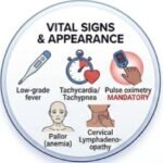 Fever is often low-grade in early disease. Tachycardia, tachypnoea, and hypoxaemia may be present in advanced disease. Cachexia or wasting, pallor from anemia of chronic disease, and lymphadenopathy (particularly cervical) are common systemic findings. Pulse oximetry is mandatory and should be recorded on room air where possible. An oxygen saturation below 92% on room air in a patient without chronic lung disease is a red flag.
For online earning ideas, freelancing, affiliate marketing, YouTube, Facebook monetization, and AI tips, visit ssthem.xyz for online earning and WhatsApp group links
Chest Examination: 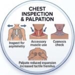 On inspection, look for asymmetry of chest movement, use of accessory muscles, and cyanosis. On palpation, reduced chest expansion and increased tactile fremitus over an area of consolidation may be detected, though these findings are operator-dependent. Dullness to percussion over a consolidated area is a classic but relatively insensitive sign. On auscultation, crackles (rales), bronchial breath sounds, and egophony are the most commonly taught signs of consolidation. However, their sensitivity is modest. The absence of crackles does not exclude TB.
Findings Suggesting Complications: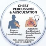 Pleural effusion may present with stony dullness, reduced breath sounds, and reduced vocal resonance. Miliary TB may present with hepatosplenomegaly, choroidal tubercles on fundoscopy, and diffuse crackles. Tuberculous meningitis may present with neck stiffness, photophobia, and altered consciousness.
Findings Suggesting Alternative Diagnoses: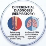 Unilateral leg swelling and pleuritic pain suggest pulmonary embolism. Bilateral basal crackles, raised jugular venous pressure, and peripheral oedema suggest heart failure. Cachexia, haemoptysis, and chronic cough suggest lung cancer. Diffuse wheeze and prolonged expiration suggest asthma or COPD exacerbation.
Red Flags That Should Not Be Ignored
The following table summarizes clinically meaningful warning signs that require urgent attention. These are not exaggerated risks but genuine indicators of potentially serious underlying pathology.
| Red Flag | Why It Matters | Possible Concern | Appropriate Action |
|---|---|---|---|
| Massive hemoptysis | Can cause airway obstruction and asphyxiation | Ruptured Rasmussen aneurysm, advanced cavitary disease | Emergency assessment, airway protection, embolization consideration |
| Severe breathlessness | Suggests extensive parenchymal involvement or pleural disease | Extensive TB, pneumothorax, pleural effusion | Urgent chest imaging, oxygen, specialist review |
| Confusion or altered consciousness | Suggests central nervous system involvement | Tuberculous meningitis | Urgent lumbar puncture, CSF analysis, empiric treatment |
| Neck stiffness with headache | Classic meningeal signs | Tuberculous meningitis | Immediate CSF examination, imaging |
| Miliary pattern on chest X-ray | Indicates hematogenous dissemination | Miliary TB | Urgent full evaluation, blood cultures, fundoscopy |
| Rapid clinical deterioration | Suggests disseminated or overwhelming infection | Disseminated TB, sepsis | Hospital admission, broad evaluation |
| Symptoms in very young children | Children are at higher risk of severe dissemination | Pediatric TB, miliary TB, TB meningitis | Urgent pediatric evaluation |
Differential Diagnosis
Tuberculosis shares clinical features with several conditions. The ability to distinguish these is a core clinical skill. The clinical reasoning behind these distinctions is not simply pattern recognition. It is about integrating the time course, the patient’s risk profile, the physical examination findings, and the initial investigation results.
| Differential Diagnosis | Similar Features | Distinguishing Features | Key Investigation |
|---|---|---|---|
| Community-acquired pneumonia | Cough, fever, productive sputum, consolidation on imaging | Acute onset, responds to antibiotics, no weight loss or night sweats | Sputum culture, response to empiric antibiotics |
| Lung malignancy | Cough, hemoptysis, weight loss, mass or cavitary lesion on imaging | Older age, smoking history, no fever, non-responsive to antibiotics | CT-guided biopsy, histopathology |
| Non-tuberculous mycobacterial (NTM) infection | Chronic cough, cavitary disease, positive AFB smear | Often underlying lung disease (COPD, bronchiectasis), slower progression | Species identification by molecular testing or culture |
| Fungal infection (histoplasmosis, aspergillosis) | Chronic cough, fever, cavitary lesions, weight loss | Endemic exposure, immunocompromised state, specific imaging features | Fungal serology, antigen testing, biopsy |
| Sarcoidosis | Hilar lymphadenopathy, pulmonary infiltrates, fatigue | Bilateral hilar adenopathy, no fever, elevated ACE levels | Biopsy showing non-caseating granulomas |
| Bronchiectasis | Chronic productive cough, hemoptysis | Long-standing history, recurrent infections, characteristic CT findings | High-resolution CT chest |
| Pleural mesothelioma | Pleural effusion, chest pain, weight loss | Asbestos exposure, pleural thickening, no fever | Pleural biopsy, histopathology |
The key to differentiating TB from these conditions is the combination of chronicity, systemic symptoms, epidemiological risk factors, and the pattern of imaging findings. TB characteristically involves the upper lobes and may show cavitation, while many mimics have different distributions or clinical courses. A patient with sudden-onset pleuritic pain and a clear chest X-ray is more likely to have a pulmonary embolism than TB. An older patient with orthopnoea, bilateral crackles, and a raised JVP is more likely to have heart failure. The diagnostic process should remain open to revision as new information emerges. It is worth remembering that respiratory and cardiac emergencies often overlap. If you have ever wondered how a heart attack differs from a cardiac arrest — and why the distinction matters in an emergency — our article on heart attack versus cardiac arrest explains the critical differences and the minutes that save a heart.
Diagnostic Tests
1. Molecular WHO-Recommended Rapid Diagnostics (mWRDs):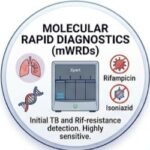 Ordered as the initial diagnostic test for all individuals with presumptive TB, replacing smear microscopy and culture as the first-line test. Xpert MTB/RIF Ultra detects M. tuberculosis complex DNA and simultaneously identifies resistance to rifampicin. It has a sensitivity of 95.3% and specificity of 94.5% with pulmonary samples. Truenat MTB/MTB Plus is a portable, chip-based test suitable for peripheral laboratories, with sensitivity of 92.9% among smear- and culture-positive samples. A positive result confirms TB. Xpert MTB/RIF Ultra can give a “trace” result, which is considered positive in children and people living with HIV. A negative result does not exclude TB, especially in paucibacillary disease. Limitations include lower sensitivity in smear-negative disease and in extrapulmonary samples. A positive NAAT result on a sputum sample generally confirms pulmonary TB. A positive result prompts treatment initiation and further drug susceptibility testing. A negative result in a patient with high clinical suspicion requires additional testing (culture, repeat molecular testing, or invasive sampling).
2. AFB Smear Microscopy:  Historically the initial test; now used as an adjunct or in settings where molecular testing is unavailable. It detects acid-fast bacilli in stained specimens. A positive smear in a patient with compatible clinical and radiological findings is highly suggestive of TB. Limitations include low sensitivity — only about 60% of culture-positive respiratory specimens are smear-positive. It cannot distinguish M. tuberculosis from non-tuberculous mycobacteria.
3. Mycobacterial Culture: 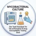 The gold standard for diagnosis and drug susceptibility testing. It grows M. tuberculosis from clinical specimens. A positive culture confirms TB. Limitations include slow turnaround — liquid culture takes 10–21 days for initial identification, and negative cultures require 6–8 weeks of incubation. Sensitivity is 80–90% depending on specimen type. False negatives can occur with inadequate specimens, prior antibiotic use, or paucibacillary disease. Culture is itself the gold standard. Positive cultures undergo species identification and drug susceptibility testing.
4. Line Probe Assay (LPA): 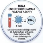 For rapid detection of drug resistance, particularly to rifampicin and isoniazid. It detects specific mutations associated with drug resistance. It is useful on smear-positive specimens or cultured isolates, especially when rifampicin resistance is suspected. Limitations include a requirement for a minimum bacillary load (approximately 10,000 bacilli/mL). Lower sensitivity for isoniazid resistance compared to rifampicin.
5. Interferon-Gamma Release Assay (IGRA):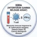 To detect TB infection (latent TB) by measuring interferon-gamma release from T-cells exposed to M. tuberculosis-specific antigens. It is useful for diagnosing latent TB infection (LTBI) and as part of the evaluation for active TB in certain contexts. Important limitation: neither IGRA nor TST can distinguish latent infection from active TB disease, and they should not be used alone for active TB diagnosis. False positives are less prone to BCG cross-reactivity than TST but can occur with certain non-tuberculous mycobacteria. False negatives can occur with immunosuppression, including HIV and TNF-α inhibitor therapy.
6. Tuberculin Skin Test (TST): 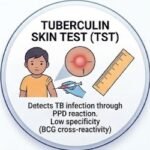 To detect TB infection through delayed-type hypersensitivity response. It measures immune response to purified protein derivative (PPD). It is useful for LTBI diagnosis, especially in children and in settings where IGRA is unavailable. Limitations include low specificity due to BCG vaccination and non-tuberculous mycobacteria cross-reactivity. Sensitivity is approximately 90% but specificity around 85%. False positives can occur with prior BCG vaccination and exposure to environmental mycobacteria. False negatives can occur with immunosuppression, malnutrition, and severe TB disease (anergy).
7. Urine Lipoarabinomannan (LAM) Assay: As a point-of-care test for active TB in people living with HIV, particularly those with advanced immunosuppression. It measures lipoarabinomannan antigen in urine. WHO strongly recommends LF-LAM to assist in the diagnosis of active TB in HIV-positive adults, adolescents, and children. Limitations include suboptimal sensitivity in HIV-negative individuals. Performance is highest in those with CD4 counts below 100 cells/μL.
8. C-Reactive Protein (CRP): As a screening tool for active TB, particularly in people living with HIV. It measures systemic inflammation. WHO recommends CRP in serum as a TB screening tool for people living with HIV. Limitations include high sensitivity (89–100%) but low specificity (0–43%). Not suitable as a standalone diagnostic test. A CRP ≥5 mg/L is recommended as the lowest threshold indicating abnormality, though ≥10 mg/L is also used.
Laboratory Tests Table
| Test | Purpose | Possible Result | Interpretation | Limitations |
|---|---|---|---|---|
| Xpert MTB/RIF Ultra | Initial diagnostic test; detects TB and rifampicin resistance | Positive (including trace), Negative, Rifampicin-resistant | Positive: TB confirmed. Trace: Consider positive in children and PLHIV; requires further testing | Lower sensitivity in smear-negative and extrapulmonary samples |
| Truenat MTB/MTB Plus | Point-of-care molecular diagnosis | Positive, Negative, Rifampicin-resistant | Positive: TB confirmed | Sensitivity lower in smear-negative samples |
| AFB Smear | Rapid presumptive diagnosis | Positive, Negative | Positive: Presumptive TB; requires confirmation | Low sensitivity (~60% of culture-positive cases) |
| Mycobacterial Culture | Gold standard for diagnosis and DST | Positive, Negative | Positive: Definitive TB diagnosis | Slow (2–8 weeks) |
| Line Probe Assay | Rapid drug resistance detection | Resistance detected, Not detected | Positive: Drug-resistant TB | Requires adequate bacillary load |
| IGRA | Detection of TB infection | Positive, Negative, Indeterminate | Positive: TB infection likely; cannot distinguish latent from active | Cannot diagnose active TB alone |
| TST | Detection of TB infection | Positive (≥5, ≥10, or ≥15 mm depending on risk), Negative | Positive: TB infection likely | Low specificity with BCG vaccination |
| Urine LAM | Point-of-care diagnosis in PLHIV | Positive, Negative | Positive: Active TB likely in PLHIV | Low sensitivity in HIV-negative individuals |
| CRP | Screening for active TB | Elevated (≥5 or ≥10 mg/L), Normal | Elevated: Increased likelihood of active TB; requires confirmatory testing | Low specificity |
Imaging Studies
Chest X-ray: Remains the first-line imaging modality. It is fast, widely available, and provides information about the presence, location, and extent of pulmonary infiltrates. Classic findings of active pulmonary TB include upper lobe consolidation, cavitation, hilar lymphadenopathy, and pleural effusion. Cavitation occurs in 10–50% of cases and is associated with higher bacillary burden. A normal chest X-ray does not rule out TB, particularly in immunocompromised patients or early disease.
CT Chest: More sensitive than X-ray for detecting small cavities, lymphadenopathy, and miliary patterns. Useful for equivocal X-ray findings, suspected complications, or pre-biopsy planning.
Ultrasound: Useful for evaluating pleural effusion and guiding thoracentesis. Can detect abdominal lymphadenopathy and ascites in abdominal TB.
MRI: Preferred modality for spinal TB (Pott’s disease) and musculoskeletal TB. Detects early bone marrow edema, synovial thickening, and soft tissue involvement.
PET-CT: Not routinely used but may help identify occult sites of extrapulmonary involvement.
If you want to understand how hypertension diagnosis can overlap with respiratory and cardiovascular risk assessment, our detailed guide to hypertension diagnosis explained with BP readings and home monitoring explains the diagnostic criteria in a clear, practical way.
Specialized and Confirmatory Testing
Bronchoscopy with Bronchoalveolar Lavage (BAL) is indicated when sputum samples are negative but clinical suspicion remains high. BAL can be tested with mWRD, culture, and cytology.
Pleural Biopsy is the most reliable method for diagnosing tuberculous pleural effusion. Thoracoscopic or blind needle biopsy can provide tissue for histopathology and culture.
Lumbar Puncture is essential for suspected tuberculous meningitis. CSF analysis typically shows lymphocytic pleocytosis, elevated protein, and low glucose. Xpert MTB/RIF Ultra on CSF has sensitivity of 50–70%.
Lymph Node Biopsy — excisional biopsy has the highest sensitivity (80–100%) for tuberculous lymphadenitis. Fine-needle aspiration is less invasive and may be useful in resource-limited settings.
Adenosine Deaminase (ADA): Elevated in tuberculous pleural, pericardial, and peritoneal effusions. Pleural fluid ADA >40 IU/L is highly suggestive of tuberculous pleuritis. Pericardial fluid ADA ≥35 U/L has sensitivity of 90% and specificity of 74% for tuberculous pericarditis.
If you want to understand why some people with significant cardiovascular disease never experience classic warning symptoms, our detailed guide to the 7 silent signs of coronary artery disease explains how the disease can progress quietly for years.
Diagnostic Criteria
There is no single universally accepted diagnostic criterion for TB. Diagnosis is established by:
1. Microbiological confirmation: Isolation of M. tuberculosis from a clinical specimen (culture or NAAT). 2. Clinical diagnosis: Compatible clinical syndrome, epidemiological risk factors, and imaging findings in the absence of microbiological confirmation, with response to anti-TB therapy. 3. Histopathological diagnosis: Granulomatous inflammation with or without acid-fast bacilli on tissue biopsy.
WHO Classification of TB: Bacteriologically confirmed TB — positive culture or NAAT. Clinically diagnosed TB — no microbiological confirmation but compatible clinical and radiological findings with decision to treat.
Drug-Resistant TB Classification: Rifampicin-resistant TB (RR-TB) — resistance to rifampicin detected. Multidrug-resistant TB (MDR-TB) — resistance to at least rifampicin and isoniazid. Extensively drug-resistant TB (XDR-TB) — resistance to rifampicin, isoniazid, fluoroquinolones, and at least one second-line injectable drug.
Diagnostic Algorithm
| Step | Action | Key Consideration |
|---|---|---|
| 1. Clinical suspicion | Cough >2 weeks, fever, night sweats, weight loss, hemoptysis | Assess symptom quality, duration, and severity |
| 2. History and examination | Assess risk factors, exposure history, severity | Older adults and immunocompromised may present atypically |
| 3. Chest X-ray | Look for upper lobe consolidation, cavitation, hilar adenopathy | Normal chest X-ray does not exclude TB |
| 4. Sputum collection | Three sputum samples including early morning specimen | Induced sputum or bronchoscopy if unable to produce |
| 5. Molecular testing | Xpert MTB/RIF Ultra or Truenat | Results within hours; detects rifampicin resistance |
| 6. AFB smear and culture | Adjunct tests; culture is gold standard | Culture takes weeks; negative result does not exclude TB |
| 7. Drug susceptibility testing | Line probe assay or phenotypic testing | Essential for drug-resistant TB |
| 8. Differential diagnosis | Exclude pneumonia, malignancy, NTM, fungal infection | Clinical context and imaging guide testing |
| 9. Determine site of care | Outpatient, ward, or intensive care unit | Based on severity and clinical judgement |
| 10. Reassess at 48–72 hours | Review response to treatment; consider treatment failure | Consider alternative or complicated diagnosis if not improving |
Severity and Staging
Radiological Classification: Minimal — lesions without demonstrable cavitation, confined to a small part of one or both lungs. Moderately advanced — disseminated lesions not exceeding the total volume of one lung; total cavitary diameter <4 cm. Far advanced — lesions more extensive than moderately advanced.
Clinical Severity: Mild — minimal symptoms, no hypoxemia, limited radiological involvement. Moderate — significant symptoms, radiological involvement, but no respiratory failure. Severe — extensive radiological disease, hypoxemia, miliary pattern, or extrapulmonary involvement.
Extensive Disease Phenotype: High semiquantitative bacterial burden on molecular testing (low cycle threshold values on Xpert), extensive disease on chest X-ray, and associated with higher relapse rates post-treatment.
Special Populations
Children: Paucibacillary disease makes microbiological confirmation difficult. WHO Treatment Decision Algorithms (TDAs) use scoring systems based on symptoms and chest X-ray findings to guide treatment initiation. Stool Xpert Ultra is a valuable non-invasive diagnostic tool in children.
Pregnant Women: Both IGRA and TST are safe during pregnancy. Chest X-ray is safe with abdominal shielding. Diagnosis may be delayed due to overlap of TB symptoms with pregnancy-related symptoms. Active TB should be treated promptly; isoniazid, rifampin, and ethambutol are considered safe in pregnancy.
Older Adults: Atypical presentation is common: absence of fever or cough, predominance of weight loss and functional decline. Higher mortality associated with delayed diagnosis. Lower cavitary lesions and more atypical infiltrates, often in lower lung zones.
Immunocompromised Patients: HIV co-infection: Higher risk of extrapulmonary and disseminated TB. Lower CD4 counts associated with atypical presentations. Urine LAM testing is particularly useful. TNF-α inhibitors: Impaired granuloma formation and negative IGRA results are common. Screening before initiation is essential. Transplant recipients: Both TST and IGRA have reduced sensitivity. Tissue biopsy is often required for diagnosis.
Patients with Kidney Disease: TST performs poorly in advanced CKD; IGRA is preferred. Screening should be risk-targeted in pre-dialysis CKD and systematic at dialysis initiation.
False Positive and False Negative Results
False Positive: A test suggests TB when the patient does not have the disease. This can occur due to: IGRA/TST — prior BCG vaccination, exposure to non-tuberculous mycobacteria, or laboratory error. Molecular tests — rare, but contamination or detection of non-viable bacilli can occur. AFB smear — non-tuberculous mycobacteria can appear acid-fast. Consequences: Unnecessary treatment, drug toxicity, psychological distress, and resource waste.
False Negative: A test suggests no TB when the patient actually has the disease. This can occur due to: AFB smear — low bacillary load, inadequate specimen, or prior antibiotic use. Molecular tests — paucibacillary disease, PCR inhibitors, or extrapulmonary samples. Culture — fastidious organisms, inadequate decontamination, or prior treatment. IGRA/TST — immunosuppression, malnutrition, or severe disease (anergy). Consequences: Delayed diagnosis, disease progression, and continued transmission.
Clinicians use pretest probability (how likely is TB based on clinical presentation?), test characteristics (sensitivity and specificity), and repeat or confirmatory testing to navigate these limitations. When clinical suspicion is high, a negative test should not automatically exclude TB — further testing with repeat or alternative methods is warranted. A normal result does not always mean the disease is absent. The most useful test is not necessarily the most advanced test. Diagnosis is a process, not a single number on a laboratory report.
10 Common Diagnostic Mistakes in Tuberculosis
- Anchoring on a single diagnosis: Once pneumonia is suspected, clinicians may fail to consider TB when the clinical course is atypical or prolonged.
- Premature diagnostic closure: Accepting a negative AFB smear as definitive exclusion of TB, particularly in paucibacillary disease or immunocompromised patients.
- Ignoring red flags: Failing to recognise hemoptysis, weight loss, or night sweats as indicators of significant disease.
- Over-relying on a single laboratory test: Using a positive IGRA alone to diagnose active TB, which it cannot do.
- Misunderstanding reference ranges: Interpreting laboratory values without considering the patient’s age, comorbidities, and clinical context.
- Ignoring pretest probability: Ordering tests indiscriminately without considering how the result will alter management.
- Failing to consider differential diagnoses: Missing malignancy, NTM infection, or fungal disease that can mimic TB.
- Inappropriate test selection: Using serology for active TB diagnosis, which is not recommended by WHO.
- Misinterpreting false-positive results: Initiating treatment for a positive test without confirmatory evidence of active disease.
- Failing to reassess an evolving patient: Not repeating tests or pursuing additional investigations when the clinical picture changes.
Hypothetical Clinical Cases
These are hypothetical educational cases and do not represent real patients.
Case 1: The Young Healthcare Worker with Persistent Cough
Presentation: A 28-year-old healthcare worker presents with a three-week history of productive cough, low-grade fever, and night sweats.
Initial Differential: Pulmonary TB, community-acquired pneumonia, acute bronchitis.
History: No prior TB. BCG vaccinated as a child. Works in an emergency department. No known TB exposure.
Examination: Temperature 37.8°C, crackles in the right upper zone. No lymphadenopathy.
Investigations: Chest X-ray shows right upper lobe consolidation with a small cavity. Sputum Xpert MTB/RIF Ultra positive, rifampicin-sensitive.
Clinical Reasoning: The subacute presentation, occupational exposure risk, and classic radiological findings strongly suggested TB. The positive mWRD confirmed the diagnosis.
Diagnosis: Pulmonary TB, rifampicin-susceptible.
Key Diagnostic Lesson: In patients with compatible symptoms and risk factors, molecular testing should be performed early. A positive mWRD on sputum is sufficient for diagnosis.
Case 2: The Immunocompromised Patient with Atypical Presentation
Presentation: A 45-year-old man with HIV (CD4 count 85 cells/mm³) presents with two weeks of fever, fatigue, and generalized lymphadenopathy. No cough.
Initial Differential: HIV-related lymphadenopathy, lymphoma, disseminated TB.
History: Not on antiretroviral therapy. No prior TB. Recent incarceration.
Examination: Temperature 38.5°C, cervical and axillary lymphadenopathy, hepatosplenomegaly.
Investigations: Chest X-ray normal. Sputum mWRD negative. Urine LAM positive. Lymph node biopsy shows caseating granulomas with acid-fast bacilli.
Clinical Reasoning: In advanced HIV, TB often presents with disseminated disease and atypical findings. A normal chest X-ray does not exclude TB. Urine LAM was valuable for rapid diagnosis, and tissue biopsy confirmed the diagnosis.
Diagnosis: Disseminated TB in advanced HIV.
Key Diagnostic Lesson: In immunocompromised patients, a high index of suspicion and a multi-modal diagnostic approach are essential. Normal chest imaging and negative sputum tests do not exclude TB.
Case 3: The Patient with Pleural Effusion
Presentation: A 35-year-old woman presents with two months of progressive dyspnea, dry cough, and weight loss.
Initial Differential: Tuberculous pleural effusion, malignancy, parapneumonic effusion.
History: Immigrated from a high-burden country five years ago. No prior TB.
Examination: Reduced breath sounds and dullness at the right base.
Investigations: Chest X-ray shows a moderate right pleural effusion. Pleural fluid analysis: lymphocytic exudate, ADA 58 IU/L, glucose low. Pleural fluid Xpert MTB/RIF Ultra positive.
Clinical Reasoning: The chronic presentation, lymphocytic exudate, and elevated ADA strongly suggested tuberculous pleuritis. The positive mWRD on pleural fluid confirmed the diagnosis.
Diagnosis: Tuberculous pleural effusion.
Key Diagnostic Lesson: In pleural TB, ADA is a useful adjunctive test, but microbiological confirmation should always be attempted. Molecular testing on pleural fluid can provide rapid confirmation.
Patient-Friendly Diagnosis Guide
What Should a Patient Expect During Diagnosis?
If your doctor suspects tuberculosis, here is what you can expect during the diagnostic process.
Consultation and history: Your doctor will ask about your symptoms — cough, fever, night sweats, weight loss — and how long you have had them. They will also ask about your medical history, medications, travel, and any contact with someone who has TB.
Physical examination: Your doctor will listen to your chest with a stethoscope, check your breathing rate, measure your oxygen level with a small device on your finger, and check your lymph nodes.
Blood tests: A blood test called an interferon-gamma release assay (IGRA) may be used to check if you have been infected with TB. This test cannot tell whether the infection is latent or active.
Sputum tests: If you have a cough, you will be asked to provide sputum samples. These are tested for TB bacteria using molecular tests (which give results in hours) and culture (which takes weeks).
Chest X-ray: A chest X-ray can show changes in your lungs that suggest TB. However, a normal X-ray does not rule out TB.
Other tests: Depending on your symptoms, you may need additional tests such as a CT scan, ultrasound, or biopsy of affected tissue.
Waiting for results: Molecular tests provide results within hours. Culture results take longer — up to 8 weeks. Your doctor may start treatment while waiting for culture results if TB is strongly suspected.
Follow-up: If you are diagnosed with TB, you will need regular follow-up to monitor your response to treatment and check for side effects. Contact tracing will be done to identify and screen anyone who may have been exposed.
Why additional tests may be needed: Not all tests are perfect. Some can produce false results. Your doctor may need to repeat or add tests to confirm the diagnosis or to rule out other conditions.
When to Seek Urgent Medical Care
Seek urgent medical assessment if you or someone you are caring for experiences any of the following:
- Coughing up large amounts of blood
- Severe difficulty breathing, or inability to speak in full sentences
- Confusion or altered mental state
- Neck stiffness with severe headache
- Chest pain that is severe or worsening
- High fever that does not improve
- Symptoms that worsen rapidly over hours
- Feeling faint or dizzy
Do not wait to see if symptoms improve. Tuberculosis can deteriorate quickly, particularly in older adults and those with underlying lung disease. Do not start, stop, or change any prescription medicine based solely on the information in this article.
25 Multiple-Choice Questions
1. A 32-year-old man presents with a 3-week history of productive cough, fever, and night sweats. He recently immigrated from a country with high TB burden. What is the most appropriate initial diagnostic test?
A. Tuberculin skin test
B. Chest X-ray and sputum Xpert MTB/RIF Ultra
C. Sputum AFB smear alone
D. Blood culture
Correct Answer: B
Explanation: WHO recommends molecular rapid diagnostics (mWRD) as the initial test for all individuals with presumptive TB. Chest X-ray complements the evaluation. TST and blood culture are not first-line diagnostic tests for active pulmonary TB.
2. A 45-year-old woman with HIV (CD4 count 75 cells/mm³) presents with fever, fatigue, and generalized lymphadenopathy. Chest X-ray is normal. Sputum mWRD is negative. Which additional test is most likely to be helpful?
A. Repeat sputum mWRD
B. Urine lipoarabinomannan (LAM) assay
C. Tuberculin skin test
D. Serum antibody test for TB
Correct Answer: B
Explanation: In PLHIV with advanced immunosuppression, urine LAM testing is recommended and can aid in the diagnosis of active TB. A normal chest X-ray and negative sputum test do not exclude TB in this population.
3. A patient has a positive interferon-gamma release assay (IGRA) but no symptoms and a normal chest X-ray. What is the most likely diagnosis?
A. Active pulmonary TB
B. Latent TB infection
C. Disseminated TB
D. Non-tuberculous mycobacterial infection
Correct Answer: B
Explanation: A positive IGRA indicates TB infection but cannot distinguish between latent and active disease. The absence of symptoms and normal chest X-ray suggest latent TB infection.
4. A sputum Xpert MTB/RIF Ultra result is reported as “trace.” What does this mean?
A. The test is invalid
B. M. tuberculosis DNA is detected at very low levels
C. The patient definitely has active TB
D. The result should be ignored
Correct Answer: B
Explanation: A trace result indicates very low bacterial load. In children and PLHIV, trace is considered positive. However, trace results may also represent non-viable bacilli, and further testing is often needed.
5. Which of the following is the gold standard for TB diagnosis?
A. AFB smear microscopy
B. Chest X-ray
C. Mycobacterial culture
D. IGRA
Correct Answer: C
Explanation: Culture remains the gold standard for TB diagnosis and drug susceptibility testing, although molecular tests are now recommended as initial diagnostic tests due to their speed.
6. A 28-year-old healthcare worker has a positive TST (15 mm induration) and a positive IGRA. She has no symptoms and a normal chest X-ray. What is the next step?
A. Start anti-TB treatment immediately
B. Diagnose active TB
C. Diagnose latent TB infection and consider treatment
D. Repeat the TST in 6 months
Correct Answer: C
Explanation: The positive tests indicate TB infection, but the absence of symptoms and normal chest X-ray suggest latent TB. Treatment for LTBI should be considered after ruling out active disease.
7. Which imaging finding is most characteristic of active pulmonary TB?
A. Lower lobe consolidation
B. Upper lobe cavitation
C. Bilateral hilar adenopathy alone
D. Pleural effusion alone
Correct Answer: B
Explanation: Upper lobe cavitation is a classic finding of active pulmonary TB. Lower lobe consolidation is more typical of bacterial pneumonia.
8. A patient with suspected tuberculous meningitis undergoes lumbar puncture. Which CSF finding is most consistent with TB meningitis?
A. Neutrophilic pleocytosis with high glucose
B. Lymphocytic pleocytosis with low glucose and high protein
C. Normal CSF analysis
D. Eosinophilic pleocytosis
Correct Answer: B
Explanation: TB meningitis typically shows lymphocytic pleocytosis, elevated protein, and low glucose. Neutrophilic pleocytosis suggests bacterial meningitis.
9. What is the recommended cut-off for pleural fluid ADA in tuberculous pleural effusion?
A. >10 IU/L
B. >20 IU/L
C. >40 IU/L
D. >100 IU/L
Correct Answer: C
Explanation: Pleural fluid ADA >40 IU/L is highly suggestive of tuberculous pleuritis, with high sensitivity and specificity.
10. A patient is diagnosed with rifampicin-resistant TB. What does this mean?
A. The patient has multidrug-resistant TB
B. The patient is resistant to rifampicin only
C. The patient is resistant to all first-line drugs
D. The patient has extensively drug-resistant TB
Correct Answer: B
Explanation: Rifampicin-resistant TB (RR-TB) indicates resistance to rifampicin. Multidrug-resistant TB (MDR-TB) requires resistance to at least rifampicin and isoniazid.
11. Which of the following is NOT recommended for active TB diagnosis?
A. Sputum mWRD
B. Mycobacterial culture
C. Serological antibody tests
D. Chest X-ray
Correct Answer: C
Explanation: WHO does not recommend serological tests for active TB diagnosis due to poor sensitivity and specificity.
12. A patient with suspected TB has a negative sputum mWRD but a chest X-ray showing upper lobe cavitation. What is the most appropriate next step?
A. Exclude TB based on the negative test
B. Obtain sputum culture and consider repeat molecular testing
C. Start empiric antibiotics only
D. Order serological testing
Correct Answer: B
Explanation: A negative mWRD does not exclude TB, especially with compatible radiological findings. Culture and repeat testing should be pursued.
13. Which population is at highest risk for false-negative IGRA results?
A. Healthy adults
B. Patients on TNF-α inhibitors
C. BCG-vaccinated individuals
D. Patients with latent TB
Correct Answer: B
Explanation: Immunosuppressive therapies, including TNF-α inhibitors, impair interferon-gamma release and can cause false-negative IGRA results.
14. What is the recommended initial diagnostic strategy for a child with suspected pulmonary TB?
A. Sputum culture alone
B. Chest X-ray and gastric aspirate or stool for Xpert Ultra
C. TST alone
D. Blood culture
Correct Answer: B
Explanation: WHO recommends molecular testing on respiratory and stool samples as the initial diagnostic strategy for children with suspected pulmonary TB.
15. A patient with TB is found to have resistance to isoniazid and rifampicin. What is this called?
A. Mono-resistant TB
B. Poly-resistant TB
C. Multidrug-resistant TB (MDR-TB)
D. Extensively drug-resistant TB (XDR-TB)
Correct Answer: C
Explanation: MDR-TB is defined as resistance to at least rifampicin and isoniazid.
16. Which of the following is a red flag requiring urgent assessment in a patient with suspected TB?
A. Mild fatigue
B. Low-grade fever
C. Massive hemoptysis
D. Occasional dry cough
Correct Answer: C
Explanation: Massive hemoptysis can cause airway obstruction and is a life-threatening emergency requiring urgent intervention.
17. A 70-year-old man presents with unexplained weight loss and fatigue but no cough or fever. Chest X-ray shows lower lobe infiltrates. What should be considered?
A. TB is unlikely because of atypical presentation
B. TB should be considered despite atypical presentation
C. The patient likely has pneumonia
D. No further testing is needed
Correct Answer: B
Explanation: Elderly patients often present atypically with TB, including absence of fever and cough. A high index of suspicion is required.
18. Which molecular test detects both TB and rifampicin resistance in under 2 hours?
A. Line probe assay
B. Xpert MTB/RIF Ultra
C. AFB smear
D. Culture
Correct Answer: B
Explanation: Xpert MTB/RIF Ultra detects M. tuberculosis and rifampicin resistance in under 2 hours.
19. A pregnant woman has a positive IGRA. What is the next step?
A. Start anti-TB treatment immediately
B. Evaluate for active TB with chest X-ray and sputum testing
C. Ignore the result until after delivery
D. Repeat IGRA in the third trimester
Correct Answer: B
Explanation: A positive IGRA indicates TB infection but not necessarily active disease. Evaluation for active TB is required before considering LTBI treatment.
20. What is the main limitation of AFB smear microscopy?
A. It is too expensive
B. It has low sensitivity and cannot distinguish M. tuberculosis from NTM
C. It requires specialized equipment
D. It takes weeks to provide results
Correct Answer: B
Explanation: AFB smear has low sensitivity (~60% of culture-positive cases) and cannot differentiate M. tuberculosis from non-tuberculous mycobacteria.
21. Which of the following is true about latent TB infection (LTBI)?
A. It is contagious
B. It causes symptoms
C. It can reactivate to active TB
D. It is diagnosed by positive culture
Correct Answer: C
Explanation: LTBI is not contagious and does not cause symptoms, but it can reactivate to active TB, especially in immunocompromised individuals.
22. A patient with suspected tuberculous pericarditis has a pericardial fluid ADA of 72 U/L. What does this suggest?
A. Bacterial pericarditis
B. Tuberculous pericarditis
C. Viral pericarditis
D. Malignant pericardial effusion
Correct Answer: B
Explanation: Pericardial fluid ADA ≥35 U/L is highly suggestive of tuberculous pericarditis, with sensitivity of 90% and specificity of 74%.
23. What is the recommended approach for drug susceptibility testing in MDR-TB?
A. Phenotypic testing only
B. Molecular testing with line probe assay and/or targeted NGS
C. No testing needed
D. Chest X-ray alone
Correct Answer: B
Explanation: WHO recommends rapid molecular testing, including line probe assays and targeted next-generation sequencing, for detecting drug resistance.
24. A patient with HIV and CD4 count <100 cells/mm³ has a negative sputum mWRD. What additional test is recommended?
A. Repeat sputum mWRD
B. Urine LF-LAM
C. Chest X-ray alone
D. No further testing
Correct Answer: B
Explanation: WHO strongly recommends using LF-LAM to assist in the diagnosis of active TB in HIV-positive adults with advanced immunosuppression.
25. Which statement about chest X-ray in TB diagnosis is correct?
A. A normal chest X-ray definitively excludes TB
B. Chest X-ray can confirm TB without microbiological testing
C. A normal chest X-ray does not exclude TB
D. Chest X-ray is the gold standard for TB diagnosis
Correct Answer: C
Explanation: A normal chest X-ray does not exclude TB, particularly in immunocompromised patients and those with early or extrapulmonary disease. Microbiological confirmation is required for definitive diagnosis.
Question. How is tuberculosis diagnosed?
Answer : TB diagnosis involves a combination of medical history, physical examination, chest X-ray, and laboratory tests. Molecular tests (such as Xpert MTB/RIF Ultra or Truenat) are the preferred initial diagnostic tests. Culture remains the gold standard for confirmation and drug susceptibility testing.
Question. What is the first test for TB?
Answer : The first test is typically a molecular rapid diagnostic test (mWRD) on sputum or other appropriate specimens, combined with a chest X-ray. In some settings, AFB smear microscopy may still be used.
Question. What blood tests are used for TB?
Answer : The interferon-gamma release assay (IGRA) is a blood test used to detect TB infection. It cannot distinguish between latent and active TB. Blood tests are not used to diagnose active TB directly.
Question. Can TB be diagnosed with a blood test?
Answer : Blood tests such as IGRA can detect TB infection but cannot diagnose active TB disease. Active TB requires microbiological confirmation from sputum or tissue samples.
Question. What does a positive TB test mean?
Answer : A positive molecular test or culture confirms active TB. A positive IGRA or TST indicates TB infection, which may be latent or active — further evaluation is needed.
Question. Can TB tests be falsely negative?
Answer : Yes. False-negative results can occur due to low bacterial load, inadequate specimens, prior antibiotic use, or immunosuppression. A negative test does not exclude TB when clinical suspicion is high.
Question. Can TB tests be falsely positive?
Answer : Yes, though less commonly with molecular tests. IGRA and TST can be falsely positive due to BCG vaccination or exposure to non-tuberculous mycobacteria.
Question. When is imaging needed for TB?
Answer : Chest X-ray is part of the initial evaluation for suspected pulmonary TB. CT may be needed for equivocal findings, complications, or extrapulmonary disease. MRI is preferred for spinal and musculoskeletal TB.
Question. When is a biopsy needed for TB?
Answer : Biopsy is often needed for extrapulmonary TB, such as lymph node, pleural, or bone involvement, when less invasive tests are inconclusive.
Question. When should a specialist be consulted?
Answer : Specialist referral is recommended for drug-resistant TB, complicated disease, extrapulmonary TB, pediatric TB, TB in pregnancy, and TB in immunocompromised patients.
Question. How long does TB diagnosis take?
Answer : Molecular tests provide results within hours. Culture results take 2–8 weeks. Diagnosis may take longer if multiple tests and procedures are needed.
Question. What is the most accurate test for TB?
Answer : Culture remains the gold standard for diagnosis. Molecular tests are highly accurate and provide rapid results but may miss paucibacillary disease.
Question. How is latent TB diagnosed?
Answer : Latent TB is diagnosed by a positive IGRA or TST in an asymptomatic person with a normal chest X-ray and no microbiological evidence of active disease.
Question. How is active TB diagnosed?
Answer : Active TB is diagnosed by microbiological confirmation (positive molecular test or culture) from a clinical specimen, supported by clinical and radiological findings.
Question. When should urgent medical care be sought?
Answer : Seek urgent care for massive hemoptysis, severe breathlessness, confusion, neck stiffness, or rapid clinical deterioration.
Latest Evidence & References
- World Health Organization. (2025). WHO consolidated guidelines on tuberculosis. Module 3: Diagnosis (2nd ed.). Geneva: WHO.
- World Health Organization. (2025, August 5). WHO operational handbook on tuberculosis: Module 3: Diagnosis.
- World Health Organization. (2025, April 16). WHO launches an update on the consolidated guidelines to diagnose tuberculosis.
- Centers for Disease Control and Prevention. (2025). Clinical Testing and Diagnosis for Tuberculosis.
- Centers for Disease Control and Prevention. (2025). Clinical and Laboratory Diagnosis for Tuberculosis.
- Centers for Disease Control and Prevention. (2026). Tuberculosis in Pregnancy.
- World Health Organization. (2026, March 9). WHO recommends near point-of-care tests, tongue swabs, and sputum pooling for TB diagnosis.
- MSD Manual Professional Edition. (2026). Tuberculosis (TB).
- Canadian Tuberculosis Standards. (2025). Chapter 3: Diagnosis of tuberculosis disease and drug-resistant tuberculosis.
- National Institutes of Health. (2024). Mycobacterium tuberculosis: Adult and Adolescent OIs.
- World Health Organization. (2025). WHO Global Tuberculosis Report 2025.
- The Lancet. (2025). Tuberculosis: advances in epidemiology, diagnostics, treatment, and prevention.
- PLOS Medicine. (2025). Diagnostic accuracy of the WHO tuberculosis treatment decision algorithms for children with presumptive tuberculosis: An individual participant data meta-analysis.
- Nature. (2025). Comparison of interferon gamma release assays and tuberculin skin test performance for diagnosing Mycobacterium tuberculosis infection.
- Scientific Reports. (2025). Diagnostic accuracy of Xpert MTB/RIF Ultra for abdominal tuberculosis.
Medical Information Disclaimer: The information provided in this article is for educational and informational purposes only and is intended for healthcare professionals, medical students, and informed general readers. It does not constitute medical advice, diagnosis, or treatment recommendations. Tuberculosis is a potentially serious condition that should be evaluated and managed by a qualified healthcare provider. Treatment decisions, including antibiotic selection, site of care, and duration of therapy, depend on the patient’s individual circumstances, comorbidities, and clinical judgment. Readers should not use this information to self-diagnose, self-treat, or make clinical decisions without appropriate professional consultation. If you have concerns about tuberculosis symptoms, seek medical attention promptly. The authors and publishers of this article do not assume any liability for any adverse effects or consequences resulting from the use or misuse of the information provided herein.
For religious knowledge, visit ssthem.com. For online earning and WhatsApp groups, visit ssthem.xyz. For health and beauty tips, visit ssthem.net.
For related articles on diagnosis and clinical reasoning, see:
- Act Fast: 7 Stroke Diagnosis Facts That Can Protect You — understanding why time-critical diagnosis matters for neurological emergencies.
- Heart Attack vs Cardiac Arrest: 10 Minutes to Save a Heart — how rapid differentiation between similar presentations can be life-saving.
- 7 Silent Signs of Coronary Artery Disease — recognizing subtle clinical clues before disease progresses.
- Hypertension Diagnosis Explained: BP Readings at Home — the importance of context and repeated measurement in diagnosis.
- What Is Diagnosis? 15 Powerful Steps to Understand — a foundational guide to clinical reasoning.
- Do You Have Diabetes? Blood Sugar Clues and Early Signs — another example of how diagnostic thresholds and pretest probability shape clinical decisions.