Hypertension Diagnosis 9 Important Tests Doctors Use to Confirm High BP
Hypertension Diagnosis Explained: BP Readings, Home Monitoring & Lab Tests
A patient arrives at a routine clinic visit with a mild headache that has lingered for several days. The triage nurse records a blood pressure of 148/92 mmHg. The physician repeats the measurement after five minutes of quiet rest: 146/90 mmHg. A diagnosis of hypertension seems straightforward. Yet the real diagnostic question has only just begun. Is this truly sustained hypertension? Could this be white coat hypertension, where office readings are elevated but daily life pressures are normal? Or could the opposite be true — masked hypertension, where office readings appear reassuring but out-of-office pressures are dangerously high?
This is a hypothetical educational case, but it reflects a daily reality in clinical practice. Hypertension is the most common modifiable risk factor for cardiovascular disease worldwide, yet its diagnosis is rarely as simple as a single elevated reading. The consequences of both overdiagnosis and underdiagnosis are substantial: unnecessary lifelong medication on one hand, and preventable stroke, heart attack, and kidney failure on the other. Accurate diagnosis requires a systematic approach that integrates multiple blood pressure measurements, out-of-office monitoring, laboratory evaluation, and careful clinical reasoning.
A normal result does not always mean the disease is absent. Diagnosis is a process, not a single number on a laboratory report.
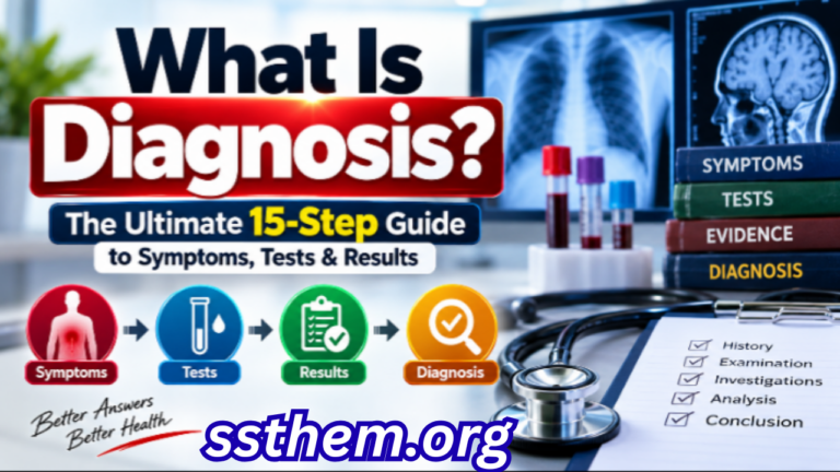
This article provides a comprehensive, evidence-based guide to hypertension diagnosis, written for medical students, residents, healthcare professionals, and informed readers who want to understand how clinicians separate true hypertension from the conditions that mimic it. If you want to understand why laboratory values can sometimes be misleading, our detailed guide to medical diagnosis explained in 15 powerful steps explains how clinicians interpret these results in context.
Key Diagnostic Facts About Hypertension
The following table summarizes the most clinically important facts about hypertension diagnosis. This is not a substitute for full clinical evaluation, but it provides a rapid reference for healthcare professionals and students.
| Diagnostic Feature | Key Fact |
|---|---|
| Disease | Hypertension (elevated arterial blood pressure) |
| Main clinical suspicion | Repeated office BP ≥130/80 mmHg (ACC/AHA) or ≥140/90 mmHg (ESC/WHO) |
| Most important symptoms | Often asymptomatic; severe cases may present with headache, visual changes, chest pain, or dyspnea |
| Important risk factors | Age, obesity, high sodium intake, physical inactivity, family history, Black ethnicity, diabetes, chronic kidney disease, sleep apnea |
| First clinical step | Standardized office blood pressure measurement with validated equipment |
| Initial investigations | Complete blood count, serum electrolytes, creatinine with eGFR, fasting glucose or HbA1c, lipid profile, TSH, urinalysis, urine albumin-to-creatinine ratio, ECG |
| Important laboratory tests | Serum creatinine/eGFR, urine albumin-to-creatinine ratio, electrolytes, glucose, lipids |
| Important imaging | Echocardiography (selective), renal artery duplex (if secondary hypertension suspected), coronary artery calcium scoring (selective) |
| Confirmatory test | Out-of-office BP monitoring (HBPM or ABPM) — essential before diagnosing hypertension |
| Important differential diagnoses | White coat hypertension, masked hypertension, secondary hypertension (renal, endocrine, vascular) |
| Major red flags | BP ≥180/120 mmHg with end-organ symptoms, sudden onset severe hypertension, hypertension in young adults (<30 years), resistant hypertension |
| Specialist referral | Suspected secondary hypertension, resistant hypertension, hypertension in pregnancy, pediatric hypertension |
| Diagnostic challenge | Distinguishing true hypertension from white coat and masked phenotypes; identifying secondary causes |
This table is a snapshot. Every parameter in it will be expanded in the dedicated sections below.
Step 1 — When Should Doctors Suspect Hypertension? 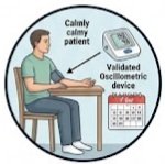
Typical Presentation: Hypertension is often called the “silent killer” because most individuals with elevated blood pressure have no symptoms. The condition is typically detected incidentally during routine health examinations, pre-employment screenings, or visits for unrelated medical concerns. When symptoms do occur, they are usually nonspecific and may include headaches (often occipital and worse in the morning), dizziness, visual disturbances, or fatigue. These symptoms are more common in severe or accelerated hypertension and should never be relied upon as the primary basis for diagnosis.
Atypical Presentation: Some patients present with symptoms of hypertensive emergencies or complications, including chest pain from myocardial ischemia, dyspnea from heart failure, focal neurological deficits from stroke, or visual loss from hypertensive retinopathy. In these cases, the diagnosis of hypertension is often made simultaneously with the complication, and immediate management is required.
Risk Factors and Epidemiological Clues: Key risk factors that increase clinical suspicion include age (prevalence increases substantially after age 40, affecting over 50% of adults over 60), obesity (particularly central adiposity), high sodium intake, low potassium intake, physical inactivity, family history of hypertension in first-degree relatives, Black ethnicity (higher prevalence and earlier onset), diabetes mellitus, chronic kidney disease, obstructive sleep apnea, metabolic syndrome, and use of medications such as nonsteroidal anti-inflammatory drugs (NSAIDs), oral contraceptives, corticosteroids, decongestants, and certain antidepressants.
Features That Increase Clinical Suspicion: Clinical suspicion for secondary hypertension should be heightened when hypertension presents with unusual features: onset before age 30, abrupt onset in previously normotensive individuals, severe or resistant hypertension, hypokalemia (suggesting primary aldosteronism), or a family history of early-onset hypertension or renal disease.
Nonspecific Findings: Many findings associated with hypertension are nonspecific, including headaches, epistaxis, and dizziness. These symptoms occur commonly in normotensive individuals and should not be attributed to hypertension without objective blood pressure confirmation.
Step 2 — Medical History 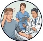
The medical history serves three purposes: to establish the diagnosis, to identify secondary causes, and to assess cardiovascular risk. Key areas to address include:
- Presenting complaint: Why is the patient seeking care? Was hypertension detected incidentally or through symptoms?
- Onset and duration: When was elevated blood pressure first noticed? Has it been documented previously?
- Progression: Have readings been stable, increasing, or fluctuating?
- Previous episodes: Any prior documentation of elevated blood pressure or diagnosis of hypertension?
- Previous diagnoses: Diabetes, kidney disease, sleep apnea, thyroid disorders, cardiovascular disease.
- Medications: Current and past use of antihypertensives, NSAIDs, oral contraceptives, corticosteroids, decongestants, stimulants, and herbal supplements.
- Allergies: Particularly to antihypertensive medications.
- Family history: Hypertension, early-onset cardiovascular disease, renal disease, endocrine tumors.
- Social history: Tobacco use, alcohol consumption, physical activity level, occupational stress.
- Dietary history: Sodium intake, potassium intake, caffeine consumption, processed food intake.
- Sleep history: Duration, quality, snoring, witnessed apnea episodes.
- Substance use: Stimulants, cocaine, amphetamines.
- Pregnancy history: Gestational hypertension or preeclampsia in previous pregnancies.
The 2025 AHA/ACC guideline emphasizes that when a new diagnosis of hypertension is suspected or confirmed, a comprehensive medical history, physical examination, and laboratory testing should be performed to inform management decisions and identify contributing causes.
For online earning ideas, freelancing, affiliate marketing, YouTube, Facebook monetization, and AI tips, visit ssthem.xyz for online earning and WhatsApp group links.
Physical Examination
Vital Signs and General Appearance: Blood pressure should be measured in both arms at the initial visit, with subsequent measurements using the arm that yields the higher reading. A difference greater than 15 mmHg between arms warrants evaluation of the upper vasculature. Heart rate, respiratory rate, oxygen saturation, and body mass index (BMI) should be documented. Waist circumference provides additional information about central adiposity.
Blood Pressure Measurement Technique: Proper technique is essential for accurate diagnosis. Avoid caffeine, exercise, and smoking for at least 30 minutes before measurement. Empty bladder. Sit quietly for at least 5 minutes before measurement. Position the patient seated with back supported, feet flat on the floor, arm supported at heart level, and legs uncrossed. Cuff bladder should encircle at least 80% of the arm circumference; width should be at least 40% of arm circumference. Use a validated oscillometric device when available. Take at least two readings 1–2 minutes apart, and average them. If readings differ by more than 5 mmHg, take additional readings. Record systolic and diastolic pressure, arm used, position, and any relevant circumstances.
Relevant Physical Findings: Fundoscopy may reveal arteriolar narrowing, arteriovenous nicking, flame hemorrhages, cotton wool spots, and papilledema, indicating hypertensive retinopathy and correlating with severity. Cardiovascular examination may reveal an S4 gallop (suggesting left ventricular hypertrophy), loud aortic component of the second heart sound, or displaced apical impulse. Diminished or delayed femoral pulses suggest coarctation of the aorta. An abdominal bruit may indicate renal artery stenosis. Thyroid examination may reveal goiter or nodules suggesting thyroid dysfunction contributing to hypertension. Skin examination may reveal striae, moon facies, and central obesity suggesting Cushing syndrome, or café-au-lait spots suggesting neurofibromatosis (pheochromocytoma association).
Findings suggesting complications include signs of heart failure (jugular venous distension, crackles, peripheral edema), stroke (focal neurological deficits), or peripheral arterial disease (diminished pulses, trophic changes).
Red Flags That Should Not Be Ignored
The following table summarizes clinically meaningful warning signs that require urgent attention. These are not exaggerated risks but genuine indicators of potentially serious underlying pathology.
| Red Flag | Why It Matters | Possible Concern | Appropriate Action |
|---|---|---|---|
| BP ≥180/120 mmHg with chest pain, dyspnea, or neurological symptoms | Indicates hypertensive emergency with acute end-organ damage | Myocardial infarction, stroke, aortic dissection, pulmonary edema | Immediate emergency department evaluation |
| Sudden onset severe hypertension in previously normotensive individual | Suggests secondary cause or acute trigger | Pheochromocytoma, renal artery embolism, drug-induced | Urgent specialist evaluation |
| Hypertension in adults younger than 30 years | Unusual for primary hypertension | Secondary hypertension (renal, endocrine, vascular) | Comprehensive secondary workup |
| Resistant hypertension (BP uncontrolled on ≥3 agents including diuretic) | Suggests secondary cause or nonadherence | Primary aldosteronism, renal artery stenosis, sleep apnea | Specialist referral, screening for secondary causes |
| Hypokalemia with hypertension | Suggests mineralocorticoid excess | Primary aldosteronism, Cushing syndrome | Aldosterone-renin ratio, further endocrine testing |
| Abdominal bruit or asymmetric kidney size | Suggests renovascular disease | Renal artery stenosis | Renal artery imaging |
| Paroxysmal headache, palpitations, diaphoresis | Classic triad of catecholamine excess | Pheochromocytoma | Plasma free metanephrines or 24-hour urine metanephrines |
Differential Diagnosis
The following table outlines the most important conditions that can resemble hypertension, along with their distinguishing features and key investigations.
| Differential Diagnosis | Similar Features | Distinguishing Features | Key Investigation |
|---|---|---|---|
| White coat hypertension | Elevated office BP readings | Normal out-of-office BP (HBPM or ABPM); prevalence 10–20% | 24-hour ABPM or 7-day HBPM |
| Masked hypertension | Normal office BP readings | Elevated out-of-office BP; prevalence 10–30% | 24-hour ABPM or HBPM |
| Primary aldosteronism | Hypertension, often resistant; hypokalemia | Low renin, elevated aldosterone; adrenal adenoma or hyperplasia | Aldosterone-renin ratio |
| Renal artery stenosis | Hypertension, often severe or resistant; abdominal bruit | Atherosclerotic or fibromuscular dysplasia; flash pulmonary edema | Duplex ultrasonography, CT angiography, MR angiography |
| Pheochromocytoma | Paroxysmal hypertension with headache, palpitations, diaphoresis | Elevated metanephrines; adrenal mass | Plasma free metanephrines, imaging |
| Cushing syndrome | Hypertension, central obesity, striae, moon facies | Elevated cortisol; ACTH-dependent or independent | Overnight dexamethasone suppression test, late-night salivary cortisol |
| Thyroid dysfunction | Hypertension, palpitations, weight changes | Abnormal TSH and free T4 | TSH, free T4 |
| Coarctation of the aorta | Hypertension in upper extremities, diminished femoral pulses | Blood pressure differential between arms and legs; echocardiographic findings | Echocardiography, CT angiography |
| Obstructive sleep apnea | Hypertension, obesity, daytime somnolence, snoring | Apnea-hypopnea index on sleep study | Polysomnography |
Clinical reasoning behind these differences hinges on the pattern of blood pressure elevation, associated symptoms, and response to targeted investigations. White coat and masked hypertension are not secondary causes but rather diagnostic phenotypes that require out-of-office confirmation to avoid misclassification.
Diagnostic Tests
Office Blood Pressure Measurement: Ordered as the initial screening and diagnostic tool for all patients. It measures brachial artery pressure at a single point in time. Useful at every clinical encounter for adults. Two or more readings ≥130/80 mmHg (ACC/AHA) or ≥140/90 mmHg (ESC/WHO) on separate occasions support diagnosis. Consistently normal readings (<120/80 mmHg) argue against it. Limitations include white coat effect, measurement error, and biological variability. Cannot diagnose hypertension alone — out-of-office confirmation is required.
Home Blood Pressure Monitoring (HBPM): Ordered to confirm diagnosis, detect white coat and masked hypertension, and monitor treatment response. It measures multiple blood pressure readings taken by the patient at home over several days. Useful before initiating treatment and for long-term monitoring. Average home BP ≥130/80 mmHg (ACC/AHA) or ≥135/85 mmHg (ESC) supports diagnosis. Requires validated device and proper technique; patient adherence varies.
24-Hour Ambulatory Blood Pressure Monitoring (ABPM): The reference standard for diagnosis of hypertension, white coat hypertension, and masked hypertension. It measures blood pressure over 24 hours, including daytime and nighttime averages. Useful when office and home readings are discordant and to assess nocturnal dipping. 24-hour average ≥130/80 mmHg; daytime average ≥135/85 mmHg; nighttime average ≥120/70 mmHg support diagnosis. Limitations include cost, availability, patient tolerance, and potential unsuitability for patients with irregular heart rhythms.
Laboratory Tests Table
| Test | Purpose | Possible Result | Interpretation | Limitations |
|---|---|---|---|---|
| Complete blood count | Assess for anemia, polycythemia | Polycythemia may sugges t sleep apnea or erythropoietin excess | Baseline; not diagnostic of hypertension | Nonspecific |
| Serum electrolytes | Assess for hypokalemia, hypercalcemia | Hypokalemia suggests primary aldosteronism; hypercalcemia suggests hyperparathyroidism | Guides secondary workup | Affected by medications, hydration |
| Serum creatinine with eGFR | Assess kidney function | Reduced eGFR indicates chronic kidney disease | Essential baseline; guides drug selection | eGFR equations vary by population |
| Fasting glucose or HbA1c | Screen for diabetes | Elevated glucose or HbA1c indicates diabetes or prediabetes | Comorbidity affecting risk and treatment | HbA1c affected by hemoglobin variants |
| Lipid profile | Assess cardiovascular risk | Elevated LDL, low HDL increase risk | Guides statin therapy decisions | Fasting status may affect results |
| Thyroid-stimulating hormone | Screen for thyroid dysfunction | Hyperthyroidism or hypothyroidism may contribute to hypertension | Treatable secondary cause | TSH may be normal in central thyroid disease |
| Urinalysis | Assess for hematuria, proteinuria | Proteinuria suggests renal disease | Indicates target organ damage | Transient proteinuria can occur |
| Urine albumin -to-creatinine ratio | Detect microalbuminuria | Elevated ratio indicates early kidney damage | Important prognostic marker | Affected by exercise, infection, hyperglycemia |
| Electrocardiogram (ECG) | Assess for left ventricular hypertrophy | LVH indicates target organ damage | Guides risk stratification | Low sensitivity for LVH compared to echocardiography |
Imaging Studies
Echocardiography: Not universally recommended for all patients with hypertension. Considered in patients with symptoms of heart failure, young patients (<18 years), or those with chronic uncontrolled hypertension to assess for left ventricular hypertrophy. It provides information on cardiac structure and function that can refine risk assessment.
Renal artery duplex ultrasonography: Recommended as the initial screening test when renovascular disease is suspected, offering a noninvasive, radiation-free assessment. Limitations include operator dependence and limited visualization in obese patients.
Coronary artery calcium scoring: May be considered for cardiovascular risk stratification in select patients, particularly those with intermediate risk where treatment decisions are uncertain.
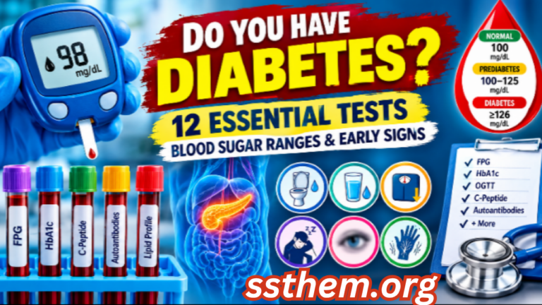
If you want to understand how blood sugar clues can signal early metabolic disease — often coexisting with hypertension — our detailed guide to diabetes blood sugar clues and early signs explains the diagnostic overlap in a clear, practical way.
Diagnostic Criteria
Hypertension diagnostic criteria vary by guideline. The 2025 AHA/ACC guideline maintains the threshold of ≥130/80 mmHg for stage 1 hypertension, a definition first introduced in 2017. The European Society of Cardiology (ESC) 2024 guideline uses ≥140/90 mmHg as the threshold for hypertension, with 120–139/70–89 mmHg classified as “elevated blood pressure”. The World Health Organization (WHO) continues to recommend ≥140/90 mmHg for diagnosis and initiation of pharmacological treatment.
| Category | ACC/AHA 2025 | ESC 2024 | WHO 2025 |
|---|---|---|---|
| Normal | <120/80 mmHg | <120/70 mmHg | <120/80 mmHg |
| Elevated | 120–129/<80 mmHg | 120–139/70–89 mmHg | 120–139/80–89 mmHg |
| Stage 1 hypertension | 130–139/80–89 mmHg | ≥140/90 mmHg | ≥140/90 mmHg |
| Stage 2 hypertension | ≥140/90 mmHg | — | — |
Diagnosis requires confirmation with out-of-office blood pressure monitoring (HBPM or ABPM) unless blood pressure is severely elevated (≥180/120 mmHg) with evidence of end-organ damage.
Diagnostic Algorithm
| Step | Action | Decision Point |
|---|---|---|
| 1. Screening | Measure office BP at routine visits | BP ≥130/80 mmHg (ACC/AHA) or ≥140/90 mmHg (ESC/WHO) |
| 2. Confirm elevated reading | Repeat office measurement on separate occasion | Persistent elevation |
| 3. Out-of-office confirmation | HBPM (7 days, twice daily) or ABPM (24 hours) | Average HBPM ≥130/80 or ABPM 24h ≥130/80 |
| 4. Assess cardiovascular risk | History, physical examination, laboratory tests | Identify risk factors, target organ damage |
| 5. Screen for secondary causes | Based on clinical clues (young age, resistant hypertension, hypokalemia, etc.) | Targeted investigations |
| 6. Classify severity | Stage 1 vs. Stage 2; presence of target organ damage | Guides treatment intensity |
| 7. Specialist referral | If secondary cause suspected, resistant hypertension, pregnancy, or pediatric | Further evaluation |
Severity and Staging
Hypertension severity is classified based on blood pressure levels and the presence of target organ damage. Stage 1 hypertension (130–139/80–89 mmHg) is associated with increased cardiovascular risk but may be managed initially with lifestyle modification in low-risk individuals. Stage 2 hypertension (≥140/90 mmHg) generally warrants pharmacological therapy in addition to lifestyle changes. Hypertensive urgency (BP ≥180/120 mmHg without end-organ damage) and hypertensive emergency (BP ≥180/120 mmHg with end-organ damage) require urgent medical attention.
Target organ damage — including left ventricular hypertrophy, chronic kidney disease, retinopathy, and vascular disease — significantly influences prognosis and treatment decisions.
Special Populations
Children and adolescents: Hypertension is defined using age-, sex-, and height-specific percentiles. Secondary causes are more common in children than adults, and referral to a pediatric specialist is recommended.
Older adults: Isolated systolic hypertension is common due to arterial stiffness. Diagnostic thresholds are the same, but treatment targets may be individualized based on frailty and comorbidities.
Pregnancy: Hypertension in pregnancy requires careful evaluation for preeclampsia and gestational hypertension. Blood pressure thresholds for treatment differ, and certain antihypertensive medications are contraindicated.
Chronic kidney disease: Hypertension is both a cause and consequence of kidney disease. Lower blood pressure targets may be appropriate, and certain drug classes (ACE inhibitors, ARBs) are preferred for renoprotection.
Diabetes mellitus: Hypertension and diabetes frequently coexist, compounding cardiovascular risk. Blood pressure targets may be lower, and ACE inhibitors or ARBs are preferred first-line agents.
False Positive and False Negative Results
False Positive: A false-positive result occurs when a test indicates hypertension when the condition is actually absent. The most common example is white coat hypertension, where office readings are elevated but out-of-office readings are normal. This affects approximately 10–20% of the population and is more common in children, older adults, and women. Overdiagnosis can lead to unnecessary medication and associated adverse effects.
False Negative: A false-negative result occurs when a test suggests normal blood pressure when hypertension is present. Masked hypertension — normal office readings but elevated out-of-office readings — affects 10–30% of adults and is associated with cardiovascular risk similar to sustained hypertension. Underdiagnosis leaves patients at risk for preventable complications.
Clinicians use pretest probability, clinical findings, and repeat or confirmatory testing to minimize these errors. Out-of-office monitoring is the cornerstone of accurate diagnosis.
The most useful test is not necessarily the most advanced test. Sometimes the most important diagnostic tool is a properly measured blood pressure reading taken at home.
Common Diagnostic Mistakes in Hypertension
- Diagnosing hypertension on a single office reading: Blood pressure varies throughout the day; confirmation with out-of-office monitoring is essential.
- Anchoring on the first elevated reading: This cognitive bias can lead to premature diagnostic closure and failure to consider white coat hypertension.
- Ignoring out-of-office monitoring: Failing to use HBPM or ABPM misses white coat and masked hypertension, leading to misclassification.
- Over-relying on symptoms: Most hypertension is asymptomatic; diagnosis should not depend on the presence of headaches or other nonspecific symptoms.
- Misunderstanding reference ranges: Blood pressure thresholds differ between guidelines; clinicians should apply the criteria appropriate to their population and setting.
- Failing to screen for secondary causes: Young age, resistant hypertension, and specific clinical clues should prompt targeted investigation.
- Inappropriate test selection: Ordering echocardiography or renal imaging in all patients rather than targeting those with clinical indications.
- Misinterpreting white coat hypertension: Assuming all patients with elevated office readings have sustained hypertension.
- Missing masked hypertension: Assuming normal office readings exclude hypertension.
- Failing to reassess: Hypertension is an evolving condition; periodic reassessment is necessary, particularly after treatment initiation or changes.
Hypothetical Clinical Cases
These are hypothetical educational cases and do not represent real patients.
Case 1: White Coat Hypertension
Presentation: A 45-year-old woman with no significant medical history presents for a routine check-up. Office blood pressure is 152/94 mmHg. She reports no symptoms. She is not taking any medications.
Initial Differential: Essential hypertension, white coat hypertension, secondary hypertension.
History: No family history of hypertension. Non-smoker, occasional alcohol. No NSAID use. No snoring or daytime somnolence.
Examination: BMI 24. Fundoscopy normal. No abdominal bruit. Peripheral pulses intact.
Investigations: HBPM over 7 days shows average home BP 118/74 mmHg. Laboratory tests including electrolytes, creatinine, glucose, and urinalysis are normal.
Test Results: Home BP average 118/74 mmHg; office BP 152/94 mmHg.
Clinical Reasoning: The discordance between office and home readings suggests white coat hypertension. Normal laboratory tests make secondary causes less likely.
Diagnosis: White coat hypertension. Lifestyle modification and annual monitoring recommended, with transition to sustained hypertension a possibility.
Key Diagnostic Lesson: Out-of-office monitoring is essential to avoid overdiagnosis and unnecessary treatment.
Case 2: Masked Hypertension
Presentation: A 52-year-old man with a history of type 2 diabetes presents for a routine visit. Office BP is 126/78 mmHg. He reports occasional morning headaches. He takes metformin but no antihypertensive medications.
Initial Differential: Normotension, masked hypertension, diabetes-related hypertension.
History: Family history of hypertension. Non-smoker. BMI 31. Reports snoring and daytime fatigue.
Examination: Waist circumference 108 cm. Fundoscopy shows mild arteriolar narrowing. ECG shows possible left ventricular hypertrophy.
Investigations: HBPM average 142/88 mmHg. Laboratory tests show HbA1c 7.2%, creatinine normal, urine albumin-to-creatinine ratio elevated.
Test Results: Home BP average 142/88 mmHg; office BP 126/78 mmHg.
Clinical Reasoning: Normal office readings with elevated home readings indicate masked hypertension. The presence of target organ damage (LVH, albuminuria) supports a diagnosis of sustained hypertension.
Diagnosis: Masked hypertension with target organ damage. Antihypertensive therapy initiated.
Key Diagnostic Lesson: Normal office blood pressure does not exclude hypertension, particularly in high-risk patients.
Case 3: Secondary Hypertension — Primary Aldosteronism
Presentation: A 38-year-old woman presents with resistant hypertension on three antihypertensive medications, including a diuretic. Office BP is 158/98 mmHg. She reports muscle cramps and fatigue.
Initial Differential: Essential hypertension, primary aldosteronism, renal artery stenosis, pheochromocytoma.
History: No family history of early-onset hypertension. No NSAID use.
Examination: BMI 27. No abdominal bruit. No cushingoid features.
Investigations: Serum potassium 3.1 mmol/L (low). Aldosterone-renin ratio elevated. Adrenal CT shows a left adrenal nodule.
Test Results: Aldosterone-renin ratio: aldosterone 28 ng/dL, renin 0.3 ng/mL/h. Adrenal CT: 1.5 cm left adrenal nodule.
Clinical Reasoning: Resistant hypertension with hypokalemia strongly suggests primary aldosteronism. The elevated aldosterone-renin ratio and adrenal nodule confirm the diagnosis.
Diagnosis: Primary aldosteronism due to aldosterone-producing adenoma. Adrenalectomy considered.
Key Diagnostic Lesson: Resistant hypertension with hypokalemia should prompt screening for primary aldosteronism.
Patient-Friendly Diagnosis Guide
What Should a Patient Expect During Diagnosis?
If your doctor suspects hypertension, the diagnostic process typically unfolds in several steps:
Consultation and history: Your doctor will ask about your medical history, medications, lifestyle, family history, and any symptoms. Be honest about all medications and supplements you take.
Physical examination: Your blood pressure will be measured in both arms. Your doctor may examine your heart, lungs, abdomen, and eyes for signs of hypertension-related complications.
Blood tests: A blood sample will be taken to check your kidney function, electrolyte levels, blood sugar, cholesterol, and thyroid function.
Urine tests: A urine sample will be checked for protein, blood, and other abnormalities that may indicate kidney damage.
Home blood pressure monitoring: You may be asked to measure your blood pressure at home twice daily for 7 days using a validated device. This provides a more accurate picture of your usual blood pressure.
24-hour ambulatory blood pressure monitoring: In some cases, you may wear a portable monitor for 24 hours that takes readings at regular intervals, including during sleep.
Imaging: If secondary hypertension is suspected, imaging studies such as ultrasound, CT, or MRI may be ordered.
Waiting for results: Some test results are available quickly, while others may take several days. Your doctor will discuss the results with you and explain what they mean.
Follow-up: If hypertension is confirmed, your doctor will discuss treatment options and schedule regular follow-up visits.
When to Seek Urgent Medical Care
Seek urgent medical care if you experience:
- Blood pressure reading ≥180/120 mmHg with chest pain, shortness of breath, severe headache, vision changes, confusion, or weakness on one side of the body
- Sudden severe headache unlike any you have experienced before
- Difficulty speaking or understanding speech
- Chest pain or pressure that radiates to the arm, neck, or jaw
- Sudden weakness or numbness in the face, arm, or leg
- Difficulty breathing or shortness of breath at rest
- Sudden vision loss or double vision
These symptoms may indicate a hypertensive emergency or a cardiovascular event requiring immediate evaluation.
1. A 50-year-old man has office blood pressure readings of 138/86 mmHg on two separate visits. According to the 2025 AHA/ACC guideline, how should this be classified?
A. Normal blood pressure
B. Elevated blood pressure
C. Stage 1 hypertension
D. Stage 2 hypertension
Correct Answer: C
Explanation: The 2025 AHA/ACC guideline defines stage 1 hypertension as systolic BP 130–139 mmHg or diastolic BP 80–89 mmHg.
2. Which of the following is the most accurate method to confirm a diagnosis of hypertension?
A. Two office readings on the same day
B. A single elevated reading with symptoms
C. 24-hour ambulatory blood pressure monitoring
D. Home blood pressure monitoring for 3 days
Correct Answer: C
Explanation: ABPM is considered the reference standard for diagnosing hypertension and detecting white coat and masked hypertension.
3. A 35-year-old woman has office BP of 150/95 mmHg but normal home readings. What is the most likely diagnosis?
A. Stage 2 hypertension
B. White coat hypertension
C. Masked hypertension
D. Secondary hypertension
Correct Answer: B
Explanation: White coat hypertension is defined as elevated office BP with normal out-of-office readings.
4. Which laboratory test is most useful for screening for primary aldosteronism?
A. Serum potassium
B. Aldosterone-renin ratio
C. Plasma metanephrines
D. Renal artery duplex
Correct Answer: B
Explanation: The aldosterone-renin ratio is the screening test of choice for primary aldosteronism.
5. A patient with resistant hypertension and hypokalemia should be screened for which secondary cause?
A. Pheochromocytoma
B. Primary aldosteronism
C. Renal artery stenosis
D. Cushing syndrome
Correct Answer: B
Explanation: Resistant hypertension with hypokalemia is a classic presentation of primary aldosteronism.
6. Which of the following is NOT a recognized risk factor for hypertension?
A. High sodium intake
B. Physical inactivity
C. Low potassium intake
D. Regular aerobic exercise
Correct Answer: D
Explanation: Regular aerobic exercise reduces blood pressure and is not a risk factor for hypertension.
7. A 28-year-old man presents with severe hypertension and abdominal bruit. Which condition should be suspected?
A. Essential hypertension
B. Renal artery stenosis
C. White coat hypertension
D. Primary aldosteronism
Correct Answer: B
Explanation: Abdominal bruit in a young patient with severe hypertension suggests renal artery stenosis.
8. What is the recommended duration for home blood pressure monitoring to confirm hypertension?
A. 1 day
B. 3 days
C. 7 days
D. 14 days
Correct Answer: C
Explanation: Guidelines recommend 7 days of home monitoring, with at least 5 days of readings, for diagnosis.
9. Which ECG finding is most suggestive of hypertensive target organ damage?
A. ST elevation
B. Left ventricular hypertrophy
C. Right axis deviation
D. Prolonged QT interval
Correct Answer: B
Explanation: Left ventricular hypertrophy is a classic ECG finding of hypertensive heart disease.
10. A patient with masked hypertension has:
A. Elevated office and out-of-office BP
B. Normal office and elevated out-of-office BP
C. Elevated office and normal out-of-office BP
D. Normal office and out-of-office BP
Correct Answer: B
Explanation: Masked hypertension is defined as normal office BP with elevated out-of-office readings.
11. Which of the following is a red flag warranting urgent evaluation?
A. BP 130/85 mmHg with mild headache
B. BP 160/100 mmHg on routine visit
C. BP 190/120 mmHg with chest pain
D. BP 145/90 mmHg in a patient with diabetes
Correct Answer: C
Explanation: BP ≥180/120 mmHg with chest pain suggests hypertensive emergency requiring immediate evaluation.
12. What is the first-line imaging test if renovascular disease is suspected?
A. CT angiography
B. Renal artery duplex ultrasonography
C. MRI
D. Renal scintigraphy
Correct Answer: B
Explanation: Duplex ultrasonography is the recommended initial noninvasive screening test for renal artery stenosis.
13. A 55-year-old woman with hypertension and diabetes should ideally be treated with which first-line drug class?
A. Beta-blocker
B. ACE inhibitor or ARB
C. Loop diuretic
D. Alpha-blocker
Correct Answer: B
Explanation: ACE inhibitors or ARBs are preferred first-line agents in hypertension with diabetes for renoprotection.
14. Which of the following statements about white coat hypertension is correct?
A. It carries no cardiovascular risk
B. It is more common in men
C. It may progress to sustained hypertension
D. It does not require monitoring
Correct Answer: C
Explanation: White coat hypertension is associated with a 3- to 4-fold increased risk of developing sustained hypertension over 7–10 years.
15. What is the most appropriate initial action for a patient with a single elevated office BP reading?
A. Start antihypertensive medication immediately
B. Repeat measurement on a separate occasion
C. Order echocardiography
D. Refer to a cardiologist
Correct Answer: B
Explanation: Diagnosis requires confirmation with repeat measurements and out-of-office monitoring.
16. Which of the following is a limitation of office blood pressure measurement?
A. It cannot detect white coat hypertension
B. It is invasive
C. It requires hospitalization
D. It measures nighttime BP
Correct Answer: A
Explanation: Office readings cannot distinguish white coat hypertension from sustained hypertension.
17. A 40-year-old man has an aldosterone-renin ratio that is elevated. What does this suggest?
A. Pheochromocytoma
B. Primary aldosteronism
C. Cushing syndrome
D. Renal artery stenosis
Correct Answer: B
Explanation: An elevated aldosterone-renin ratio is the hallmark of primary aldosteronism.
18. Which of the following is NOT part of the initial laboratory workup for hypertension?
A. Complete blood count
B. Serum creatinine with eGFR
C. Coronary angiography
D. Urinalysis
Correct Answer: C
Explanation: Coronary angiography is not part of routine initial hypertension workup.
19. A patient has an average 24-hour ABPM of 128/76 mmHg. What is the interpretation?
A. Normal
B. Hypertension
C. White coat hypertension
D. Masked hypertension
Correct Answer: B
Explanation: The 24-hour ABPM threshold for hypertension is ≥130/80 mmHg (ACC/AHA). A value of 128/76 is borderline but may be considered elevated in some contexts.
20. Which of the following best describes the relationship between hypertension and symptoms?
A. Most patients have headaches
B. Symptoms are reliable for diagnosis
C. Most patients are asymptomatic
D. Epistaxis is a specific symptom
Correct Answer: C
Explanation: Hypertension is typically asymptomatic and is often detected incidentally.
21. A patient with hypertension and hypokalemia should be evaluated for:
A. Diabetes
B. Primary aldosteronism
C. Thyroid disease
D. Pheochromocytoma
Correct Answer: B
Explanation: Hypokalemia in hypertension suggests mineralocorticoid excess, most commonly primary aldosteronism.
22. What is the purpose of echocardiography in hypertension evaluation?
A. To diagnose hypertension
B. To assess for left ventricular hypertrophy and cardiac function
C. To measure renal artery flow
D. To screen for pheochromocytoma
Correct Answer: B
Explanation: Echocardiography assesses cardiac structure and function, particularly for LVH.
23. Which of the following is a limitation of home blood pressure monitoring?
A. It cannot detect masked hypertension
B. It requires validated device and proper technique
C. It is invasive
D. It measures nighttime BP
Correct Answer: B
Explanation: HBPM requires a validated device and correct technique to be reliable.
24. A 60-year-old man has BP 170/100 mmHg on three antihypertensive medications including a diuretic. What is this called?
A. White coat hypertension
B. Resistant hypertension
C. Masked hypertension
D. Hypertensive urgency
Correct Answer: B
Explanation: Resistant hypertension is defined as BP above goal despite three agents including a diuretic.
25. Which guideline maintains a diagnostic threshold of ≥130/80 mmHg for hypertension?
A. WHO
B. ESC
C. AHA/ACC
D. NICE
Correct Answer: C
Explanation: The AHA/ACC guideline defines hypertension as ≥130/80 mmHg, while ESC and WHO use ≥140/90 mmHg.
Question. How is hypertension diagnosed?
Answer : Hypertension is diagnosed through repeated blood pressure measurements. Office readings are confirmed with home blood pressure monitoring (HBPM) or 24-hour ambulatory blood pressure monitoring (ABPM). Diagnosis requires average readings at or above guideline thresholds.
Question. What is the first test for hypertension?
Answer : The first test is a properly measured office blood pressure reading using a validated device. If elevated, out-of-office monitoring is recommended to confirm the diagnosis.
Question. What blood tests are used to diagnose hypertension?
Answer : Initial blood tests include complete blood count, serum electrolytes, creatinine with eGFR, fasting glucose or HbA1c, lipid profile, thyroid-stimulating hormone, and urinalysis. These tests assess cardiovascular risk and screen for secondary causes.
Question. Can hypertension be diagnosed without a specific test?
Answer : Hypertension is diagnosed based on blood pressure measurements, not a single test. Out-of-office monitoring is essential to confirm the diagnosis.
Question. What does a positive test mean?
Answer : A positive result in hypertension diagnosis means that blood pressure readings are consistently at or above the diagnostic threshold, confirmed by out-of-office monitoring.
Question. Can diagnostic tests be falsely negative?
Answer : Yes. Masked hypertension is a form of false-negative result, where office readings are normal but out-of-office readings are elevated. This affects 10–30% of adults.
Question. Can diagnostic tests be falsely positive?
Answer : Yes. White coat hypertension is a form of false-positive result, where office readings are elevated but out-of-office readings are normal. This affects 10–20% of the population.
Question. When is imaging needed?
Answer : Imaging is needed when secondary hypertension is suspected. Examples include renal artery duplex for renovascular disease, adrenal imaging for primary aldosteronism, and echocardiography for cardiac involvement.
Question. When is biopsy needed?
Answer : Biopsy is rarely needed for hypertension diagnosis. It may be considered in rare cases of renal parenchymal disease when other tests are inconclusive.
Question. When should a specialist be consulted?
Answer : Specialist referral is recommended for suspected secondary hypertension, resistant hypertension, hypertension in pregnancy, pediatric hypertension, and complex cases requiring advanced evaluation.
Question. When should urgent medical care be sought?
Answer : Seek urgent care if blood pressure is ≥180/120 mmHg with chest pain, shortness of breath, severe headache, vision changes, confusion, or focal neurological deficits.
Question. Can you have high blood pressure without symptoms?
Answer : Yes. Most people with hypertension have no symptoms. This is why it is called the “silent killer” and why routine screening is important.
Question. What blood pressure readings indicate hypertension?
Answer : According to the 2025 AHA/ACC guideline, stage 1 hypertension is 130–139/80–89 mmHg and stage 2 is ≥140/90 mmHg. The ESC and WHO use ≥140/90 mmHg as the threshold.
Question. How to measure blood pressure correctly?
Answer : Sit quietly for 5 minutes with back supported and feet flat. Use a validated upper-arm cuff. Avoid caffeine, exercise, and smoking for 30 minutes before. Take at least two readings 1–2 minutes apart and average them.
Question. What is the difference between white coat and masked hypertension?
Answer : White coat hypertension is elevated office BP with normal out-of-office readings. Masked hypertension is normal office BP with elevated out-of-office readings. Both require out-of-office monitoring for accurate diagnosis.
Latest Evidence & References
- Whelton PK, Carey RM, Aronow WS, et al. 2017 ACC/AHA/AAPA/ABC/ACPM/AGS/APhA/ASH/ASPC/NMA/PCNA Guideline for the Prevention, Detection, Evaluation, and Management of High Blood Pressure in Adults. Hypertension. 2018;71(6):e13-e115.
- McEvoy JW, McCarthy CP, Bruno RM, et al. 2024 ESC Guidelines for the management of elevated blood pressure and hypertension. European Heart Journal. 2024;45(38):3912-4018.
- 2025 AHA/ACC/AANP/AAPA/ABC/ACCP/ACPM/AGS/AMA/ASPC/NMA/PCNA/SGIM Guideline for the Prevention, Detection, Evaluation, and Management of High Blood Pressure in Adults. Journal of the American College of Cardiology. 2025.
- Mancia G, Kreutz R, Brunström M, et al. 2023 ESH Guidelines for the management of arterial hypertension. Journal of Hypertension. 2023;41(12):1874-2071.
- World Health Organization. Guideline for the pharmacological treatment of hypertension in adults. Geneva: WHO; 2021 (updated 2025).
- National Institute for Health and Care Excellence. Hypertension in adults: diagnosis and management. NICE guideline NG136. 2019 (updated 2024).
- Unger T, Borghi C, Charchar F, et al. 2020 International Society of Hypertension Global Hypertension Practice Guidelines. Hypertension. 2020;75(6):1334-1357.
- Kario K, Hoshide S, Mogi M, et al. The Japanese Society of Hypertension Guidelines for the Management of Hypertension (JSH2025). Hypertension Research. 2026;49(1):1-100.
- Stergiou GS, Palatini P, Parati G, et al. 2021 European Society of Hypertension practice guidelines for office and out-of-office blood pressure measurement. Journal of Hypertension. 2021;39(7):1293-1302.
- Parati G, Stergiou GS, Bilo G, et al. Home blood pressure monitoring: methodology, clinical relevance and practical application. Journal of Hypertension. 2021;39(9):1742-1762.
- 2025 AHA/ACC Hypertension Guideline: Out-of-Office BP Monitoring. Journal of the American College of Cardiology. 2025.
- Muntner P, Shimbo D, Carey RM, et al. Measurement of Blood Pressure in Humans: A Scientific Statement from the American Heart Association. Hypertension. 2019;73(5):e35-e66.
- Carey RM, Calhoun DA, Bakris GL, et al. Resistant Hypertension: Detection, Evaluation, and Management. Hypertension. 2018;72(5):e53-e90.
- Funder JW, Carey RM, Mantero F, et al. The Management of Primary Aldosteronism: Case Detection, Diagnosis, and Treatment. Journal of Clinical Endocrinology & Metabolism. 2016;101(5):1889-1916.
- Lenders JW, Duh QY, Eisenhofer G, et al. Pheochromocytoma and Paraganglioma: An Endocrine Society Clinical Practice Guideline. Journal of Clinical Endocrinology & Metabolism. 2014;99(6):1915-1942.
Medical Information Disclaimer: The information provided in this article is for educational and informational purposes only and is intended for healthcare professionals, medical students, and informed general readers. It does not constitute medical advice, diagnosis, or treatment recommendations. Hypertension is a medical condition that should be evaluated and managed by a qualified healthcare provider. Treatment decisions, including medication selection, dose adjustment, and monitoring, depend on the patient’s individual circumstances, comorbidities, and clinical judgment. Readers should not use this information to self-diagnose, self-treat, or make clinical decisions without appropriate professional consultation. If you have concerns about your blood pressure or cardiovascular health, seek evaluation from a qualified healthcare professional. The authors and publishers of this article do not assume any liability for any adverse effects or consequences resulting from the use or misuse of the information provided herein.
For authentic and engaging information about the world’s different religions, visit ssthem.com for religious studies and discover well-organized, informative content.



3 Comments