Act FAST: 7 Stroke Diagnosis Facts That Can Protect You and Your Family
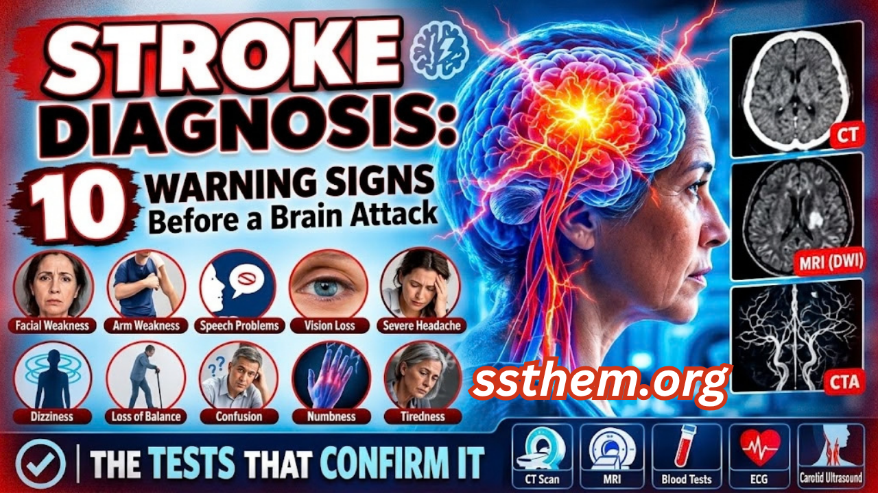
Stroke Diagnosis 10 Warning Signs That May Appear Before a Brain Attack — And the Tests That Confirm It
A 67-year-old woman arrives at the emergency department with a symptom that appears routine: sudden weakness in her right arm while pouring tea. By the time the triage nurse sees her, the weakness has almost completely resolved. Her blood pressure is mildly elevated. The initial non-contrast CT scan of her brain is normal. It would be easy to conclude that this was nothing serious. Yet the real diagnostic question has only just begun: what evidence can separate a transient ischemic attack from a resolved stroke, a seizure with postictal paresis, a hemiplegic migraine, or a dozen other conditions that can mimic cerebrovascular disease? The answer lies not in a single test, but in a disciplined diagnostic process that unfolds under extreme time pressure. Stroke diagnosis is a race against time — every minute of delay may cost approximately 1.9 million neurons. The purpose of this article is not to replace clinical judgment but to explain, at a professor-of-medicine level, exactly how clinicians think when they suspect a stroke.
This is a hypothetical educational case, but it reflects a daily reality in clinical practice. Stroke — whether ischemic, hemorrhagic, or transient ischemic attack — remains a leading cause of death and disability worldwide, yet its diagnosis is rarely as simple as a single normal scan. The consequences of both overdiagnosis and underdiagnosis are substantial: unnecessary thrombolysis on one hand, and preventable brain damage, disability, and death on the other. Accurate diagnosis requires a systematic approach that integrates clinical history, neurological examination, neuroimaging, laboratory testing, and careful clinical reasoning.
A normal CT scan does not always mean the disease is absent. 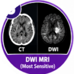 Diagnosis is a process, not a single image on a screen.
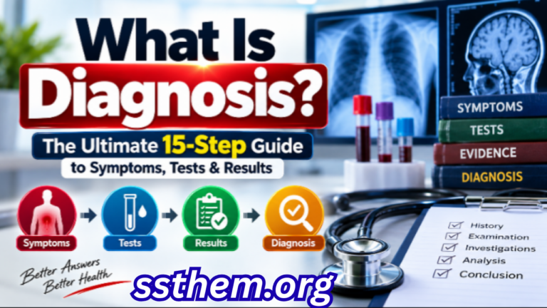
This article provides a comprehensive, evidence-based guide to Stroke Diagnosis, written for medical students, residents, healthcare professionals, and informed readers who want to understand how clinicians separate true stroke from the conditions that mimic it. If you want to understand why laboratory values and imaging findings can sometimes be misleading, our detailed guide to medical diagnosis explained in 15 powerful steps explains how clinicians interpret these results in context.
Key Diagnostic Facts About Stroke
The following table summarizes the most clinically important facts about stroke diagnosis. This is not a substitute for full clinical evaluation, but it provides a rapid reference for healthcare professionals and students.
| Diagnostic Feature | Key Fact |
|---|---|
| Disease | Stroke (ischemic, hemorrhagic, and transient ischemic attack) |
| Main clinical suspicion | Sudden-onset focal neurological deficit: facial weakness, arm or leg weakness, speech disturbance, visual loss, or ataxia |
| Most important symptoms | Unilateral weakness or numbness, dysphasia, dysarthria, hemianopia, diplopia, ataxia, sudden severe headache (hemorrhagic) |
| Important risk factors | Hypertension, atrial fibrillation, diabetes mellitus, smoking, hyperlipidemia, prior TIA/stroke, carotid stenosis, age >55, family history |
| First clinical step | ABC assessment, blood glucose, and validated stroke screening tool (FAST, BE-FAST, or ROSIER) |
| Initial investigations | Non-contrast CT head (NCCT) within 20–25 minutes of arrival; blood glucose, CBC, coagulation profile, electrolytes, renal function |
| Important laboratory tests | Glucose (to exclude hypoglycemia), CBC (to exclude thrombocytopenia before thrombolysis), INR/PTT (to exclude coagulopathy), creatinine (for contrast imaging) |
| Important imaging | NCCT (first-line to exclude hemorrhage); CT angiography (CTA) for large vessel occlusion; CT perfusion (CTP) or MRI with diffusion-weighted imaging (DWI) for tissue-based selection |
| Confirmatory test | No single “confirmatory” test. Diagnosis is clinical + imaging. DWI-MRI is the most sensitive for acute ischemia. |
| Important differential diagnoses | Hypoglycemia, seizure with Todd paresis, migraine with aura, Bell’s palsy, functional neurological disorder, hypertensive encephalopathy, central vertigo (HINTS examination) |
| Major red flags | Sudden severe headache (“thunderclap”), decreased consciousness, vomiting, neck stiffness, focal deficit with anticoagulant use, recent head trauma |
| Specialist referral | Immediate stroke team activation; neurology or stroke specialist assessment; neurosurgery for large hemorrhagic stroke or hydrocephalus |
| Diagnostic challenge | Up to one-third of suspected strokes are ultimately diagnosed as stroke mimics; NCCT can be normal in hyperacute ischemia; posterior circulation strokes are frequently missed. |
This table is a snapshot. Every parameter in it will be expanded in the dedicated sections below.
Step 1 — When Should Doctors Suspect a Stroke?
Typical Presentation: 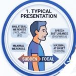 Stroke is defined by the World Health Organization as “a clinical syndrome consisting of rapidly developing clinical signs of focal (or global) disturbance of cerebral function, lasting more than 24 hours or leading to death, with no apparent cause other than that of vascular origin.” The diagnosis is fundamentally clinical. Imaging confirms the type and location but does not make the diagnosis in isolation. The classic presentation is sudden-onset, focal, non-progressive neurological deficit. The most common symptoms are unilateral weakness or paralysis of the face, arm, or leg; dysphasia (language disturbance); ataxia; visual disturbance; and sensory loss. The deficit is maximal at onset — it does not “spread” gradually over hours as a tumor or demyelinating lesion might.
Early Warning Signs: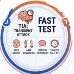 A transient ischemic attack (TIA) is a transient episode of neurological dysfunction caused by focal brain, spinal cord, or retinal ischemia, without acute infarction on imaging. Symptoms typically last minutes and usually resolve within one hour, but even very short episodes are clinically significant because the risk of recurrent stroke is highest in the first 7 days. The FAST test (Face, Arm, Speech, Time) has a pooled sensitivity of approximately 79–95% but variable specificity of 52–84%, meaning it is good at catching strokes but also triggers evaluation for many non-strokes.
Atypical Presentation: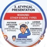 Posterior circulation strokes may present with vertigo, diplopia, dysarthria, dysphagia, or crossed sensory findings rather than the classic hemiparesis. Small vessel lacunar strokes may present with pure motor or pure sensory deficits without cortical signs. Hemorrhagic stroke may present with sudden severe headache, vomiting, and decreased consciousness. In young adults (age <50), arterial dissection, patent foramen ovale, hypercoagulable states, and vasculitis become more prominent. Approximately 10% of ischemic strokes occur in young adults.
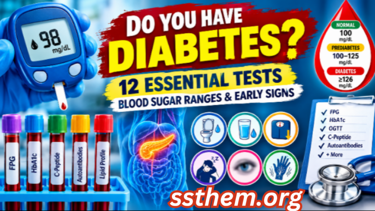
Nonspecific Findings: 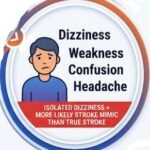 Dizziness, generalized weakness, confusion, and headache are common in both stroke and non-stroke conditions. Isolated dizziness without other focal findings is more often a stroke mimic than a true stroke. If you want to understand how another major vascular risk factor — diabetes — can quietly damage blood vessels for years before producing symptoms, our detailed guide to diabetes blood sugar clues and early signs explains the diagnostic overlap in a clear, practical way.
Step 2 — Medical History
The history is the single most powerful diagnostic tool in stroke. Because treatment decisions depend on time of onset, the first question is always: When was the patient last known well? This defines the treatment window. A focused but thorough history should elicit the following:
- Presenting complaint: What exactly happened? Was the onset sudden or gradual? Which body parts are involved? Has the deficit improved, worsened, or fluctuated?
- Onset and duration: The exact time of symptom onset or last known well is critical. Wake-up strokes and unwitnessed strokes require imaging-based selection for reperfusion therapy.
- Progression: Stroke deficits are typically maximal at onset. Stepwise progression suggests repeated emboli or expanding hematoma. Gradual progression over hours to days suggests tumor, abscess, or demyelination.
- Associated symptoms: Headache (sudden severe headache suggests hemorrhage or venous thrombosis), vomiting (suggests raised intracranial pressure or hemorrhage), neck pain (suggests arterial dissection), visual disturbance, and loss of consciousness.
- Previous episodes: Prior TIA, stroke, atrial fibrillation, myocardial infarction, or transient monocular blindness (amaurosis fugax) all increase the probability of a vascular event.
- Medications: Anticoagulants (warfarin, direct oral anticoagulants) increase the risk of hemorrhagic stroke and contraindicate thrombolysis. Antiplatelets, antihypertensives, and statins are relevant to both risk and management.
- Family history: Premature cardiovascular disease, stroke in first-degree relatives, and known genetic disorders (e.g., CADASIL, Fabry disease) may be relevant, particularly in young stroke patients.
- Social history: Smoking, alcohol use, recreational drug use (cocaine, amphetamines), and occupation. Cocaine and amphetamines are potent vasoconstrictors and can cause both ischemic and hemorrhagic stroke.
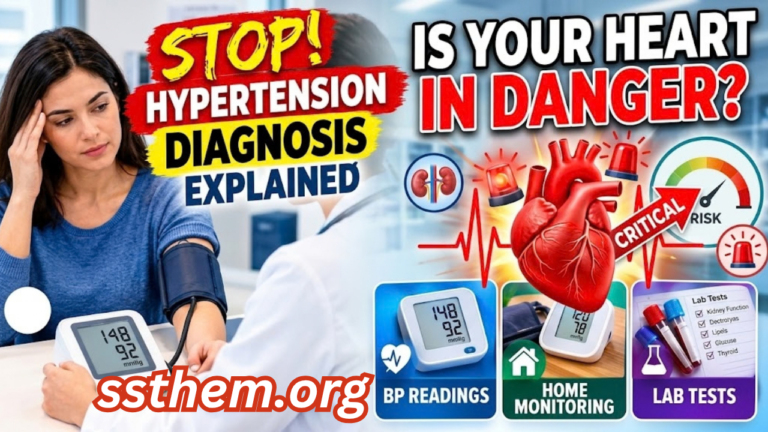
The history is a hypothesis-generating exercise. A patient who describes “the worst headache of my life” while gardening is a different diagnostic problem from a patient who woke up with a drooping face. If you want to understand how hypertension — the single most important modifiable risk factor for stroke — is diagnosed and monitored, our detailed guide to hypertension diagnosis explained with BP readings and home monitoring explains the diagnostic criteria in a clear, practical way.
Physical Examination
Vital Signs and General Appearance: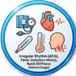 Blood pressure (both arms), heart rate and rhythm (irregularly irregular suggests atrial fibrillation), respiratory rate, oxygen saturation, and temperature. Fever may suggest infective endocarditis or another infectious mimic. Level of consciousness (Glasgow Coma Scale), neck stiffness (subarachnoid hemorrhage or meningitis), and signs of trauma should be assessed.
For online earning ideas, freelancing, affiliate marketing, YouTube, Facebook monetization, and AI tips, visit ssthem.xyz for online earning and WhatsApp group links
Neurological Examination: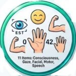 The National Institutes of Health Stroke Scale (NIHSS) is the standard severity assessment tool. It scores 11 items including level of consciousness, gaze, visual fields, facial palsy, motor function of arms and legs, ataxia, sensory loss, language, dysarthria, and extinction. Scores range from 0 (no symptoms) to 42 (severe stroke). A score of 1–4 is considered minor stroke, 5–15 moderate, 16–20 moderate-to-severe, and 21–42 severe.
Specific Diagnostic Signs: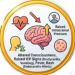 In stroke, facial palsy often spares the forehead (upper motor neuron pattern) because the forehead receives bilateral cortical innervation. In Bell’s palsy, the entire half of the face including the forehead is weak (lower motor neuron pattern). Dysphasia (language disturbance — wrong words, inability to comprehend) suggests cortical involvement. Dysarthria (slurred speech due to motor weakness) can occur with brainstem or cerebellar lesions. Homonymous hemianopia suggests a retrochiasmal lesion, often parietal or occipital. Truncal ataxia with vertigo and nystagmus should prompt the HINTS examination (Head Impulse, Nystagmus, Test of Skew) to distinguish central from peripheral causes.
Findings Suggesting Complications: Decreased consciousness suggests large hemispheric stroke with edema, brainstem involvement, or hemorrhagic transformation. Vomiting and bradycardia suggest raised intracranial pressure. Fever, rash, meningismus, fluctuating deficits, and bilateral symptoms all point away from a single vascular territory stroke.
Red Flags That Should Not Be Ignored
The following table summarizes clinically meaningful warning signs that require urgent attention. These are not exaggerated risks but genuine indicators of potentially serious underlying pathology.
| Red Flag | Why It Matters | Possible Concern | Appropriate Action |
|---|---|---|---|
| Thunderclap headache | Sudden severe headache peaks in seconds | Subarachnoid hemorrhage | Urgent NCCT; if negative and suspicion high, lumbar puncture or CTA |
| Decreased consciousness | Suggests large territory involvement or hemorrhage | Large hemispheric infarct, brainstem stroke, intracerebral hemorrhage | Immediate stroke team activation, airway assessment, urgent imaging |
| Vomiting with focal deficit | Suggests raised intracranial pressure or hemorrhage | Hemorrhagic stroke, cerebellar stroke with hydrocephalus | Urgent NCCT, neurosurgical consultation |
| Focal deficit on anticoagulant | High risk of hemorrhagic transformation | Intracerebral hemorrhage | Urgent NCCT, coagulation studies, reversal agents if indicated |
| Recent head trauma | Trauma can cause subdural hematoma mimicking stroke | Subdural or epidural hematoma | Urgent NCCT |
| Neck pain with neurological deficit | Suggests arterial dissection | Carotid or vertebral artery dissection | CTA or MRA of neck vessels |
Differential Diagnosis
The differential diagnosis of stroke is broad. Up to one-third of patients initially suspected of having a stroke are ultimately diagnosed with a stroke mimic. The most common mimics are seizures with postictal paresis (Todd’s paresis), migraine with aura, hypoglycemia, Bell’s palsy, and functional neurological disorder.
| Differential Diagnosis | Similar Features | Distinguishing Features | Key Investigation |
|---|---|---|---|
| Hypoglycemia | Focal weakness, confusion, altered consciousness | Corrects rapidly with glucose; history of diabetes; diaphoresis | Bedside glucose |
| Seizure with Todd paresis | Postictal focal weakness, confusion | Witnessed seizure, postictal confusion, no vascular risk factors; weakness resolves over minutes to hours | History, EEG, glucose |
| Migraine with aura | Visual disturbance, sensory changes, dysphasia | Gradual spread of symptoms over minutes; positive phenomena (scintillating scotoma, paresthesias); headache history; younger age | Clinical history; imaging usually normal |
| Bell’s palsy | Isolated facial weakness | Involves forehead; no limb weakness; no dysphasia | Clinical examination |
| Functional neurological disorder | Variable weakness, sensory loss | Inconsistent examination, Hoover’s sign, give-way weakness, distractibility | Clinical examination; imaging normal |
| Central vertigo (posterior circulation stroke) | Vertigo, nystagmus, ataxia | HINTS examination suggests central cause; other brainstem signs | MRI with DWI, HINTS examination |
| Brain tumor | Progressive focal deficit, seizure | Gradual onset over days to weeks; headache, papilledema | MRI with contrast |
| Subdural hematoma | Focal deficit, altered consciousness | History of trauma; gradual onset; elderly or anticoagulated | NCCT |
The clinical reasoning behind these distinctions is not memorization. It is pattern recognition. A young woman with a headache and transient visual disturbance is more likely to have migraine. An elderly man with sudden right arm weakness and atrial fibrillation has stroke until proven otherwise. A patient with isolated facial weakness that includes the forehead and no other deficits likely has Bell’s palsy. It is worth remembering that stroke and heart disease share many risk factors and sometimes present in overlapping ways. If you have ever wondered how a heart attack differs from a cardiac arrest — and why the distinction matters in an emergency — our article on heart attack versus cardiac arrest explains the critical differences and the minutes that save a heart.
Diagnostic Tests
1. Non-Contrast CT Head (NCCT): 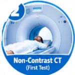 Ordered as the first-line imaging test in all suspected acute stroke. It is fast, widely available, and reliably excludes intracranial hemorrhage, which must be ruled out before thrombolysis. It measures X-ray attenuation, which reflects tissue density. Acute blood appears hyperdense (bright). Early ischemic changes — loss of gray-white differentiation, sulcal effacement — may be subtle or absent in the first hours. A hyperdense vessel sign (suggesting thrombus), loss of insular ribbon, obscured lentiform nucleus, or ASPECTS ≤7 in a patient with clinical stroke syndrome supports the diagnosis. A completely normal NCCT in the hyperacute phase does not exclude ischemic stroke. NCCT is relatively insensitive for hyperacute ischemia, particularly in the posterior fossa. A normal CT does not rule out stroke. As one clinical review noted, CT is “very inaccurate in identifying posterior circulation strokes,” and an important adverse effect of a normal CT is false reassurance.
2. CT Angiography (CTA):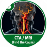 Identifies large vessel occlusion (LVO), which may qualify the patient for mechanical thrombectomy. The 2026 AHA/ASA guidelines recommend emergent CT/CTA if MRI/MRA is not available immediately (within 25 minutes). CTA visualizes the cervical and intracranial arteries with intravenous contrast. It can show occlusion, stenosis, dissection, and aneurysm. It is used in all patients with suspected large vessel occlusion who may be candidates for endovascular thrombectomy, including the 6–24 hour window. It requires intravenous contrast (risk of allergy, nephropathy). Not all hospitals have 24/7 CTA capability.
3. MRI with Diffusion-Weighted Imaging (DWI):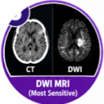 DWI is the most sensitive sequence for acute ischemia. It detects restricted diffusion within minutes of symptom onset, reflecting cytotoxic edema. It measures the random motion of water molecules. In ischemic tissue, water movement is restricted, producing a bright signal on DWI and a dark signal on the apparent diffusion coefficient (ADC) map. When the diagnosis is uncertain, when posterior circulation stroke is suspected, when the patient presents late, or when TIA is suspected but the clinician wants to exclude infarction, MRI is the test of choice. Restricted diffusion in a vascular territory consistent with the clinical deficit confirms acute ischemic stroke. MRI is less widely available than CT and takes longer to acquire. It is contraindicated in patients with certain metallic implants.
4. CT Perfusion (CTP): CTP measures cerebral blood flow, blood volume, and mean transit time. It identifies the ischemic core (irreversibly damaged tissue) and penumbra (at-risk but salvageable tissue). In the 4.5–9 hour window or wake-up stroke, CTP-based selection identifies patients who may benefit from reperfusion despite presenting late. CTP involves radiation and contrast. It requires post-processing software. False-negative CTP can occur: in one study, 118 patients had normal acute CTP but confirmed infarct on follow-up imaging.
5. Blood Tests:  Blood tests in acute stroke serve three purposes: to exclude mimics (glucose), to assess eligibility for thrombolysis (CBC, coagulation), and to identify risk factors (HbA1c, lipids). Hypoglycemia is a common stroke mimic and must be excluded immediately. Thrombocytopenia (platelet count <100,000/µL) is a contraindication to thrombolysis. INR, PTT, and thrombin time assess coagulopathy. Patients on warfarin with INR >1.7 are generally excluded from thrombolysis. Creatinine is needed before contrast imaging. Sodium abnormalities can mimic stroke. Cardiac troponin assesses for concurrent myocardial injury or cardioembolic source. A normal blood test does not mean the patient is not having a stroke. The diagnosis remains clinical and imaging-based.
Laboratory Tests Table
| Test | Purpose | Possible Result | Interpretation | Limitations |
|---|---|---|---|---|
| NCCT head | Exclude hemorrhage; assess early ischemia | Hyperdense vessel, loss of gray-white differentiation, ASPECTS ≤7 | Supports ischemic stroke; excludes hemorrhage | Normal in hyperacute phase; poor for posterior fossa |
| CTA | Identify LVO; assess neck and intracranial vessels | Occlusion, stenosis, dissection | Guides thrombectomy decision | Contrast risks; availability |
| MRI DWI | Most sensitive for acute ischemia | Restricted diffusion | Confirms acute infarct | Availability; time; contraindications |
| Glucose | Exclude hypoglycemia mimic | <70 mg/dL (varies) | Hypoglycemia causes reversible focal deficits | Must be checked immediately |
| CBC | Assess platelet count | <100,000/µL | Contraindicates thrombolysis | Stress leukocytosis is nonspecific |
| INR/PTT | Assess coagulopathy | INR >1.7 | Generally contraindicates thrombolysis | Warfarin variability; DOACs may not affect INR |
Imaging Studies
Non-Contrast CT Head: Remains the first-line imaging modality. It is fast, widely available, and reliably excludes hemorrhage. Early ischemic changes may be subtle or absent in the first hours. A normal CT does not exclude ischemic stroke.
CT Angiography: Identifies large vessel occlusion, which may qualify the patient for mechanical thrombectomy. It visualizes the cervical and intracranial arteries with intravenous contrast. It can show occlusion, stenosis, dissection, and aneurysm.
MRI with DWI: The most sensitive sequence for acute ischemia. It detects restricted diffusion within minutes of symptom onset. It is the test of choice when the diagnosis is uncertain, when posterior circulation stroke is suspected, when the patient presents late, or when TIA is suspected but the clinician wants to exclude infarction.
CT Perfusion: Measures cerebral blood flow, blood volume, and mean transit time. It identifies the ischemic core and penumbra. In the 4.5–9 hour window or wake-up stroke, CTP-based selection identifies patients who may benefit from reperfusion despite presenting late.
If you want to understand how hypertension diagnosis can overlap with stroke risk assessment, our detailed guide to hypertension diagnosis explained with BP readings and home monitoring explains the diagnostic criteria in a clear, practical way.
Specialized and Confirmatory Testing
Digital Subtraction Angiography (DSA) remains the reference standard for anatomical assessment of the cerebral vasculature. It is used when non-invasive imaging is inconclusive or when endovascular intervention is planned.
Transcranial Doppler (TCD) assesses cerebral blood flow velocity and can detect stenosis, occlusion, and emboli. It is non-invasive and can be performed at the bedside.
Carotid Ultrasound assesses the extracranial carotid arteries for stenosis or plaque. It is used in the workup of TIA and ischemic stroke to identify treatable carotid disease.
Echocardiography identifies cardioembolic sources such as atrial thrombus, valvular vegetation, or patent foramen ovale. It is recommended in all patients with cryptogenic stroke, particularly younger patients.
If you want to understand why some people with significant cardiovascular disease never experience classic warning symptoms, our detailed guide to the 7 silent signs of coronary artery disease explains how the disease can progress quietly for years.
Diagnostic Criteria
There is no single universally accepted diagnostic criterion for stroke. Diagnosis is made by:
1. Clinical presentation: Sudden-onset focal neurological deficit consistent with a vascular territory. 2. Imaging evidence: Infarction on CT or MRI, or exclusion of hemorrhage with clinical syndrome consistent with ischemia. 3. Exclusion of mimics: Glucose, seizure, migraine, and other conditions.
For TIA, the definition is tissue-based: a transient episode of neurological dysfunction caused by focal brain, spinal cord, or retinal ischemia, without acute infarction. Symptoms usually resolve within one hour, and DWI shows no restricted diffusion. For hemorrhagic stroke, diagnosis requires imaging evidence of intraparenchymal or subarachnoid blood on NCCT or MRI. The TOAST classification is used for etiological subtyping of ischemic stroke: large-artery atherosclerosis, cardioembolism, small-vessel occlusion, other determined etiology, and undetermined etiology. This classification is not a diagnostic criterion but guides secondary prevention.
Diagnostic Algorithm
| Step | Action | Key Consideration |
|---|---|---|
| 1. Symptom recognition | Sudden focal neurological deficit; FAST/BE-FAST screening | Assess symptom quality, duration, and time of onset |
| 2. ABC assessment + glucose | Check airway, breathing, circulation; bedside glucose | Hypoglycemia is a common and reversible mimic |
| 3. Urgent NCCT (target <25 minutes) | Exclude hemorrhage; assess early ischemic changes | Normal CT does not exclude ischemic stroke |
| 4. CTA ± CTP or MRI DWI | Identify large vessel occlusion; assess core and penumbra | Guides thrombectomy and thrombolysis decisions |
| 5. ECG and cardiac monitoring | Identify atrial fibrillation or other arrhythmias | Cardioembolic source guides secondary prevention |
| 6. NIHSS severity assessment | Score stroke severity; guide treatment intensity | Weighted toward anterior circulation; less sensitive for posterior stroke |
| 7. Differential diagnosis | Exclude seizure, migraine, hypoglycemia, Bell’s palsy, functional disorder | Clinical context and imaging guide testing |
| 8. Etiology workup | Echocardiography, carotid ultrasound, Holter monitoring, thrombophilia screen | Guides secondary prevention |
| 9. Specialist referral | Neurology or stroke specialist assessment | Reperfusion vs. medical therapy |
| 10. Follow-up | Secondary prevention, rehabilitation, risk factor management | Ongoing assessment |
Severity and Staging
Stroke severity is measured by the NIHSS. Scores range from 0 to 42: 1–4: Minor stroke. 5–15: Moderate stroke. 16–20: Moderate-to-severe stroke. 21–42: Severe stroke.
The ASPECTS score assesses early ischemic change on NCCT. A score of 10 indicates no visible ischemic changes in the MCA territory. Each point is subtracted for early ischemic changes in specific regions. ASPECTS ≥7 generally indicates a favorable profile for reperfusion, though the 2026 guidelines expand thrombectomy eligibility to selected patients with ASPECTS 3–5. The modified Rankin Scale (mRS) measures functional outcome and is used to assess prestroke disability and poststroke prognosis.
Special Populations
Women: Women have unique risk factors including pregnancy, preeclampsia, oral contraceptive use (especially with migraine with aura), and hormone replacement therapy. Autoimmune conditions such as lupus and antiphospholipid syndrome are more common in women. Diagnosis follows the same principles, but the differential diagnosis is broader.
Young adults (age <50): Approximately 10% of ischemic strokes occur in this group. Arterial dissection, patent foramen ovale, hypercoagulable states, and recreational drug use are more prominent. Stroke mimics are also more common in young patients. A high index of suspicion and thorough vascular imaging are essential.
Older adults: Older adults may present atypically with confusion, falls, or decreased mobility rather than classic focal deficits. Comorbidities and polypharmacy complicate diagnosis and treatment. Frailty and prestroke functional status influence treatment decisions.
Pediatric stroke: The 2026 AHA/ASA guidelines provide the first detailed recommendations for diagnosing and treating stroke in children. Emergent brain and vascular imaging with MRI/MRA is recommended to identify large vessel occlusion and differentiate ischemic stroke from hemorrhage or mimics. If MRI is not available within 25 minutes, CT/CTA is reasonable.
Pregnancy: Stroke risk is increased during pregnancy and the postpartum period. Imaging should not be withheld when clinically indicated; MRI without gadolinium is preferred, but CT with shielding may be necessary. Management requires multidisciplinary input.
False Positive and False Negative Results
False Positive: A test suggests stroke when no stroke is present. Examples include: NCCT showing an old infarct that is mistaken for acute stroke; CTA showing a stenosis that is not the cause of the current deficit; clinical screening tools (FAST) triggering stroke alerts in patients with migraine or seizure. False positives matter because they lead to unnecessary thrombolysis (risk of hemorrhage) and resource utilization.
False Negative: A test does not show stroke when stroke is present. Examples include: normal NCCT in hyperacute ischemic stroke (common); normal DWI in very small brainstem strokes; normal CTP despite confirmed infarct on follow-up. False negatives matter because they delay treatment and may lead to missed opportunities for reperfusion.
Clinicians use pretest probability (how likely is stroke based on clinical presentation?), test characteristics (sensitivity and specificity), and repeat or confirmatory testing to navigate these limitations. A normal CT in a patient with a classic clinical stroke syndrome is not reassuring — it is expected, and the next step is CTA/MRI. A normal result does not always mean the disease is absent. The most useful test is not necessarily the most advanced test. Diagnosis is a process, not a single image on a screen.
10 Common Diagnostic Mistakes in Stroke
- Anchoring on a normal CT: A normal NCCT does not exclude stroke. In hyperacute ischemia, the scan is often normal.
- Premature diagnostic closure: Diagnosing “dizziness” or “confusion” without considering posterior circulation stroke.
- Ignoring the time of onset: Treatment eligibility depends entirely on when the patient was last known well.
- Over-relying on the NIHSS: The NIHSS is weighted toward anterior circulation strokes and may underestimate posterior circulation events.
- Misunderstanding the FAST test: FAST has high sensitivity but low specificity; a positive FAST does not confirm stroke, and a negative FAST does not exclude it.
- Failing to check glucose: Hypoglycemia is a common and reversible stroke mimic.
- Not considering stroke mimics: Seizure, migraine, Bell’s palsy, and functional disorders are frequently mistaken for stroke.
- Inappropriate use of the HINTS examination: HINTS is validated for acute vertigo with nystagmus, not for all dizzy patients.
- Missing arterial dissection: Young patients with headache and neck pain may have carotid or vertebral dissection.
- Failing to reassess: Stroke is dynamic. A patient who improves may still have a stroke; a patient who worsens may have hemorrhagic transformation or expanding edema.
Hypothetical Clinical Cases
These are hypothetical educational cases and do not represent real patients.
Case 1: The Resolved Deficit
Presentation: A 58-year-old man presents with a 20-minute episode of right arm weakness and difficulty finding words. Symptoms have completely resolved by the time he arrives. He has a history of hypertension and smokes.
Initial Differential: TIA, seizure with Todd paresis, migraine with aura, hypoglycemia.
History: No headache, no seizure activity, no prior similar episodes. Glucose is normal. He takes no medications.
Examination: Normal neurological examination. Blood pressure 158/92 mmHg. Irregularly irregular pulse.
Investigations: NCCT normal. ECG shows atrial fibrillation. MRI DWI shows a small area of restricted diffusion in the left frontal lobe.
Clinical Reasoning: The clinical episode was transient, but DWI shows infarction. This is a stroke, not a TIA, because there is evidence of acute infarct on imaging. The atrial fibrillation is the likely cardioembolic source.
Diagnosis: Acute ischemic stroke (cardioembolic, likely atrial fibrillation).
Key Diagnostic Lesson: DWI-MRI reclassifies many clinically transient events as strokes. The absence of symptoms does not mean the absence of infarction.
Case 2: The Dizzy Patient
Presentation: A 72-year-old woman presents with sudden-onset vertigo, nausea, and unsteadiness. She has a history of hypertension and diabetes.
Initial Differential: Benign paroxysmal positional vertigo (BPPV), vestibular neuritis, posterior circulation stroke.
History: The vertigo is constant, not positional. She has diplopia and mild dysarthria. No hearing loss.
Examination: Horizontal nystagmus that changes direction with gaze. Head impulse test is normal. Test of skew shows vertical misalignment. These are central HINTS signs.
Investigations: NCCT is normal (expected in posterior circulation stroke). MRI DWI shows acute infarction in the right cerebellar hemisphere.
Clinical Reasoning: The HINTS examination correctly identified a central cause of vertigo. NCCT is insensitive for posterior circulation stroke, and MRI is the definitive test.
Diagnosis: Acute ischemic stroke, posterior circulation (cerebellar).
Key Diagnostic Lesson: In acute vertigo, the HINTS examination is more sensitive than early MRI for distinguishing central from peripheral causes. A normal CT does not exclude posterior circulation stroke.
Case 3: The Young Patient with Headache
Presentation: A 34-year-old woman presents with severe left-sided headache and right arm weakness that began 2 hours ago. She has a history of migraine with aura.
Initial Differential: Migraine with aura, ischemic stroke, arterial dissection.
History: The headache is unlike her usual migraine — it is more severe and continuous. She reports neck pain on the left side. No recent trauma.
Examination: Right arm weakness (4/5), left-sided neck tenderness. No visual aura.
Investigations: NCCT normal. CTA shows left internal carotid artery dissection with intimal flap.
Clinical Reasoning: The combination of headache, neck pain, and focal deficit in a young patient should prompt vascular imaging. Migraine history can be a distractor that leads to diagnostic error.
Diagnosis: Ischemic stroke secondary to left internal carotid artery dissection.
Key Diagnostic Lesson: In young patients with headache and focal deficits, arterial dissection must be excluded. A history of migraine does not protect against stroke and may paradoxically increase risk.
Patient-Friendly Diagnosis Guide
What Should a Patient Expect During Diagnosis?
If you or someone you know is suspected of having a stroke, here is what typically happens in the emergency department. A nurse or doctor will check your breathing, circulation, and blood sugar. They will ask when your symptoms started — this is the most important question. A doctor will test your strength, sensation, speech, vision, and coordination. This is scored using the NIH Stroke Scale.
A small blood sample will be taken to check your sugar, blood count, clotting, and kidney function. You will have a CT scan of your head. This is quick and tells doctors whether there is bleeding in the brain. If the CT is normal, it does not mean you are not having a stroke — it means there is no bleeding. Depending on the results, you may need a CT angiogram (which looks at blood vessels) or an MRI (which is more sensitive for detecting stroke).
Some results are available within minutes; others take longer. Doctors may start treatment before all results are back if the clinical picture is clear. After the acute phase, you may need more tests to find the cause of the stroke — such as heart monitoring, echocardiography, or blood tests for clotting disorders. Why additional tests? Not all strokes look the same on the first scan. Some strokes are small, some are in the back of the brain, and some are transient. Doctors use a combination of tests to build a complete picture.
When to Seek Urgent Medical Care
Call emergency services immediately if you or someone else experiences:
- Sudden weakness or numbness of the face, arm, or leg, especially on one side
- Sudden confusion, trouble speaking, or difficulty understanding speech
- Sudden trouble seeing in one or both eyes
- Sudden trouble walking, dizziness, loss of balance, or lack of coordination
- Sudden severe headache with no known cause
Do not wait to see if symptoms improve. Time is brain. Treatments for ischemic stroke are most effective when given within 4.5 hours of symptom onset, and some patients may benefit from thrombectomy up to 24 hours after onset. Do not drive yourself to the hospital. Call emergency services so that treatment can begin en route. Do not take aspirin unless instructed. If the stroke is hemorrhagic, aspirin can worsen bleeding.
1. A 72-year-old man presents with sudden right arm weakness and difficulty speaking. His symptoms started 90 minutes ago. What is the most appropriate first imaging test?
A. MRI with DWI
B. Non-contrast CT head
C. CT angiography
D. Carotid ultrasound
Correct Answer: B
Explanation: NCCT is the first-line imaging test to exclude hemorrhage before thrombolysis. It is fast and widely available.
2. A patient’s non-contrast CT is normal 45 minutes after the onset of left-sided weakness. What does this mean?
A. Stroke is excluded
B. Hemorrhage is excluded
C. The patient has a TIA
D. MRI is unnecessary
Correct Answer: B
Explanation: A normal NCCT in the hyperacute phase excludes hemorrhage but does not exclude ischemic stroke.
3. Which of the following is the most common stroke mimic?
A. Brain tumor
B. Seizure with postictal paresis
C. Subdural hematoma
D. Encephalitis
Correct Answer: B
Explanation: Seizures with Todd paresis are among the most common stroke mimics.
4. A patient presents with isolated facial weakness that involves the forehead. What is the most likely diagnosis?
A. Ischemic stroke
B. Bell’s palsy
C. Hemorrhagic stroke
D. TIA
Correct Answer: B
Explanation: Bell’s palsy is a lower motor neuron lesion that affects the entire half of the face, including the forehead.
5. Which score is used to assess early ischemic changes on non-contrast CT?
A. NIHSS
B. ASPECTS
C. ABCD2
D. mRS
Correct Answer: B
Explanation: ASPECTS is a 10-point scale that quantifies early ischemic changes in the MCA territory on NCCT.
6. A patient with atrial fibrillation presents with sudden right-sided weakness. What is the most likely stroke mechanism?
A. Large artery atherosclerosis
B. Cardioembolism
C. Small vessel occlusion
D. Arterial dissection
Correct Answer: B
Explanation: Atrial fibrillation is a major risk factor for cardioembolic stroke.
7. Which blood test must be checked immediately in any patient with suspected stroke?
A. Troponin
B. Glucose
C. HbA1c
D. Lipid panel
Correct Answer: B
Explanation: Hypoglycemia is a common and reversible stroke mimic.
8. A patient has an INR of 2.5 and presents with acute ischemic stroke symptoms within 2 hours of onset. What is the most appropriate management?
A. Intravenous alteplase
B. Mechanical thrombectomy evaluation
C. Aspirin 300 mg
D. Observation
Correct Answer: B
Explanation: INR >1.7 generally contraindicates intravenous thrombolysis. However, the patient may still be a candidate for mechanical thrombectomy.
9. What is the definition of TIA?
A. Neurological deficit lasting less than 24 hours
B. Transient neurological dysfunction without infarction on imaging
C. Neurological deficit that resolves within 1 hour
D. Stroke with complete recovery
Correct Answer: B
Explanation: The definition of TIA is now tissue-based: a transient episode of neurological dysfunction caused by focal ischemia without acute infarction on imaging.
10. A 34-year-old woman presents with severe headache and right arm weakness. She has a history of migraine. What diagnosis must be excluded?
A. Migraine with aura
B. Arterial dissection
C. Functional neurological disorder
D. Bell’s palsy
Correct Answer: B
Explanation: Arterial dissection is a leading cause of stroke in young adults.
11. Which imaging modality is most sensitive for acute ischemic stroke within the first hour?
A. Non-contrast CT
B. CT angiography
C. MRI with DWI
D. Carotid ultrasound
Correct Answer: C
Explanation: DWI detects restricted diffusion within minutes of ischemia onset.
12. A patient has a NIHSS score of 18. How would you classify the stroke severity?
A. Minor
B. Moderate
C. Moderate-to-severe
D. Severe
Correct Answer: C
Explanation: NIHSS 16–20 is classified as moderate-to-severe stroke.
13. Which of the following is a false-negative result in stroke diagnosis?
A. Normal NCCT in hyperacute ischemic stroke
B. Hyperdense MCA sign on NCCT
C. Restricted diffusion on DWI
D. Elevated INR
Correct Answer: A
Explanation: A normal NCCT in the first hours of ischemic stroke is a false-negative result because the scan does not show the infarct, yet stroke is present.
14. A patient presents with vertigo, nystagmus, and normal head impulse test. What is the most likely diagnosis?
A. Vestibular neuritis
B. BPPV
C. Posterior circulation stroke
D. Meniere’s disease
Correct Answer: C
Explanation: A normal head impulse test in a patient with acute vertigo and nystagmus suggests a central cause, such as posterior circulation stroke.
15. What is the primary purpose of CTA in acute stroke?
A. Exclude hemorrhage
B. Identify large vessel occlusion
C. Assess brain perfusion
D. Detect old infarcts
Correct Answer: B
Explanation: CTA identifies large vessel occlusion, which may qualify the patient for mechanical thrombectomy.
16. Which of the following is a contraindication to intravenous thrombolysis?
A. Blood glucose 120 mg/dL
B. Platelet count 80,000/µL
C. NIHSS score of 6
D. Blood pressure 160/90 mmHg
Correct Answer: B
Explanation: Thrombocytopenia (platelet count <100,000/µL) is a contraindication to thrombolysis due to bleeding risk.
17. A patient’s CT perfusion shows a large ischemic core with minimal penumbra. What does this suggest?
A. Good candidate for reperfusion
B. Poor candidate for reperfusion
C. Definite hemorrhage
D. TIA
Correct Answer: B
Explanation: A large core with minimal penumbra suggests irreversible damage with little salvageable tissue.
18. Which of the following is true about the FAST test?
A. It has high specificity
B. It has high sensitivity but variable specificity
C. It confirms stroke diagnosis
D. It replaces imaging
Correct Answer: B
Explanation: FAST has good sensitivity but variable specificity. It is a screening tool, not a diagnostic test.
19. A patient with suspected stroke has a normal NCCT and normal CTA. What is the next most appropriate step?
A. Discharge home
B. MRI with DWI
C. Lumbar puncture
D. EEG
Correct Answer: B
Explanation: MRI with DWI is the most sensitive test for acute ischemia, particularly in posterior circulation strokes.
20. Which of the following is a common diagnostic error in stroke?
A. Checking glucose immediately
B. Performing NCCT within 25 minutes
C. Anchoring on a normal CT scan
D. Using the NIHSS
Correct Answer: C
Explanation: Anchoring on a normal CT scan is a common error. A normal CT does not exclude ischemic stroke.
21. A patient on warfarin presents with sudden severe headache and vomiting. What is the most likely diagnosis?
A. Ischemic stroke
B. Hemorrhagic stroke
C. TIA
D. Migraine
Correct Answer: B
Explanation: Sudden severe headache, vomiting, and anticoagulant use strongly suggest hemorrhagic stroke.
22. What is the time window for intravenous thrombolysis in acute ischemic stroke?
A. 1 hour
B. 3 hours
C. 4.5 hours
D. 12 hours
Correct Answer: C
Explanation: Intravenous thrombolysis is generally recommended within 4.5 hours of symptom onset.
23. Which of the following distinguishes hemorrhagic stroke from ischemic stroke on NCCT?
A. Loss of gray-white differentiation
B. Hyperdense area within the brain parenchyma
C. Sulcal effacement
D. Hyperdense MCA sign
Correct Answer: B
Explanation: Acute blood appears hyperdense (bright) on NCCT.
24. A patient has a transient episode of right arm weakness that resolves within 30 minutes. MRI DWI shows no restricted diffusion. What is the diagnosis?
A. Ischemic stroke
B. TIA
C. Hemorrhagic stroke
D. Seizure
Correct Answer: B
Explanation: TIA is defined as transient neurological dysfunction without infarction on imaging.
25. Which of the following is the most important question to ask in a patient with suspected stroke?
A. What medications do you take?
B. When were you last known well?
C. Do you have a family history of stroke?
D. Have you had a recent infection?
Correct Answer: B
Explanation: The time of symptom onset or last known well determines eligibility for thrombolysis and thrombectomy.
Question. How is a stroke diagnosed?
Answer : Stroke is diagnosed through clinical assessment (sudden focal neurological deficit) combined with brain imaging, usually non-contrast CT first to exclude hemorrhage, followed by CTA or MRI to confirm ischemia and identify the affected vessel.
Question. What is the first test for a stroke?
Answer : Non-contrast CT of the head is the first test. It excludes hemorrhage and assesses for early ischemic changes. It is fast and available in most emergency departments.
Question. What blood tests are used in stroke diagnosis?
Answer : Glucose (to exclude hypoglycemia), CBC (platelet count), coagulation profile (INR, PTT), renal function, and electrolytes. These tests help exclude mimics and assess treatment eligibility.
Question. Can a blood test detect a stroke?
Answer : No blood test currently confirms or excludes stroke with sufficient accuracy. Research on biomarkers such as miRNA-125b and GFAP is ongoing, but imaging remains essential.
Question. Can a stroke be missed on CT?
Answer : Yes. NCCT is often normal in the first hours of ischemic stroke, especially in the posterior circulation. A normal CT does not exclude stroke.
Question. What is the difference between stroke and TIA?
Answer : Stroke involves infarction on imaging or persistent symptoms beyond 24 hours. TIA involves transient symptoms without infarction on DWI-MRI.
Question. What is the FAST stroke test?
Answer : FAST stands for Face, Arm, Speech, Time. It is a screening tool to help the public and prehospital providers recognize stroke symptoms. It has high sensitivity but variable specificity.
Question. How do doctors diagnose a hemorrhagic stroke?
Answer : Hemorrhagic stroke is diagnosed by NCCT showing hyperdense blood within the brain parenchyma or subarachnoid space. Further imaging (CTA or MRI) may identify the underlying cause.
Question. How is stroke severity measured?
Answer : The NIH Stroke Scale (NIHSS) measures severity. Scores range from 0 (no symptoms) to 42 (severe stroke). Scores of 1–4 are minor, 5–15 moderate, 16–20 moderate-to-severe, and 21–42 severe.
Question. Can an MRI detect a stroke days later?
Answer : Yes. MRI with DWI is most sensitive in the first hours to days. T2-FLAIR sequences become increasingly sensitive for subacute infarction after 24–48 hours.
Question. How is a silent stroke diagnosed?
Answer : A silent stroke is discovered incidentally on imaging performed for another reason. It shows an old infarct without corresponding clinical symptoms. It still indicates cerebrovascular disease and warrants risk factor assessment.
Question. When should you go to the ER for stroke symptoms?
Answer : Call emergency services immediately for any sudden focal neurological deficit — weakness, numbness, speech difficulty, visual loss, or severe headache. Do not wait to see if symptoms improve.
Question. What is the 3-hour window for stroke treatment?
Answer : The 3-hour window was the original time frame for intravenous thrombolysis. Current guidelines recommend treatment within 4.5 hours, and selected patients may benefit from thrombectomy up to 24 hours.
Question. How do doctors distinguish stroke from Bell’s palsy?
Answer : Bell’s palsy causes isolated facial weakness that includes the forehead. Stroke typically spares the forehead and may involve the arm, leg, or speech.
Question. How is stroke diagnosed in young adults?
Answer : The same principles apply, but the differential diagnosis is broader. Arterial dissection, patent foramen ovale, and hypercoagulable states are more common. Vascular imaging is essential.
Latest Evidence & References
- American Heart Association/American Stroke Association. 2026 Guideline for the Early Management of Patients With Acute Ischemic Stroke. Stroke. 2026. The guideline emphasizes imaging-based selection for reperfusion therapy and includes the first formal pediatric stroke recommendations.
- National Institute for Health and Care Excellence (NICE). Stroke and transient ischaemic attack in over 16s: diagnosis and initial management (NG128). Updated guidance emphasizes rapid recognition, immediate referral to specialist stroke services, and urgent brain imaging.
- Best Practice BMJ. Ischaemic stroke: diagnosis and initial management. Last evidence review June 2026. Defines stroke as a clinical diagnosis based on signs and symptoms, with NCCT to exclude hemorrhage and DWI-MRI as the most sensitive test for acute ischemia.
- Best Practice BMJ. Transient ischaemic attack (TIA). Last reviewed December 2025. Defines TIA as a tissue-based diagnosis: transient neurological dysfunction without acute infarction on imaging.
- Powers WJ, Rabinstein AA, Ackerson T, et al. Guidelines for the Early Management of Patients With Acute Ischemic Stroke: 2026 Update. Stroke. 2026. Key recommendations include NCCT or MRI within 20–25 minutes of arrival, CTA for large vessel occlusion, and perfusion imaging for late-window selection.
- Gonzalez NR, Prabhakaran S, Zachrison KS, et al. Response to Request for Modification of the 2026 AHA/ASA Acute Ischemic Stroke Guidelines: Endovascular Thrombectomy Imaging Selection in the 6- to 24-Hour Window. Stroke. 2026. Affirms the guideline recommendation for EVT in the 6–24 hour window with ASPECTS ≥6 and NIHSS ≥6.
- Zhang et al. Meta-analysis of endovascular thrombectomy in late windows. 2026. Pooled data from 16 studies and 12,199 patients supporting EVT in the 6–24 hour window.
- Best Practice BMJ. Transient ischaemic attack: diagnostic investigations. Recommends blood glucose, full blood count, platelet count, prothrombin time, INR, and partial thromboplastin time as first-line investigations.
- Diagnostic accuracy and post-reperfusion kinetics of serum miRNA-125b in hyperacute ischemic stroke. Irish Journal of Medical Science. 2026. Prospective case-control study showing miRNA-125b AUC of 0.910, 95% sensitivity, and 90% specificity for AIS at ED admission.
- Best Practice BMJ. Ischaemic stroke: key diagnostic factors. Lists unilateral weakness or paralysis, dysphasia, ataxia, visual disturbance, and sensory loss as the main diagnostic features.
Medical Information Disclaimer: The information provided in this article is for educational and informational purposes only and is intended for healthcare professionals, medical students, and informed general readers. It does not constitute medical advice, diagnosis, or treatment recommendations. Stroke is a medical emergency that should be evaluated and managed by a qualified healthcare provider. Treatment decisions, including thrombolysis, thrombectomy, and secondary prevention, depend on the patient’s individual circumstances, comorbidities, and clinical judgment. Readers should not use this information to self-diagnose, self-treat, or make clinical decisions without appropriate professional consultation. If you have concerns about stroke symptoms, seek emergency medical attention immediately. The authors and publishers of this article do not assume any liability for any adverse effects or consequences resulting from the use or misuse of the information provided herein.
For additional health and wellness resources, you may also find evidence-based articles on ssthem.net for health and beauty tips helpful. For religious knowledge and spiritual guidance, visit ssthem.com for religious knowledge.