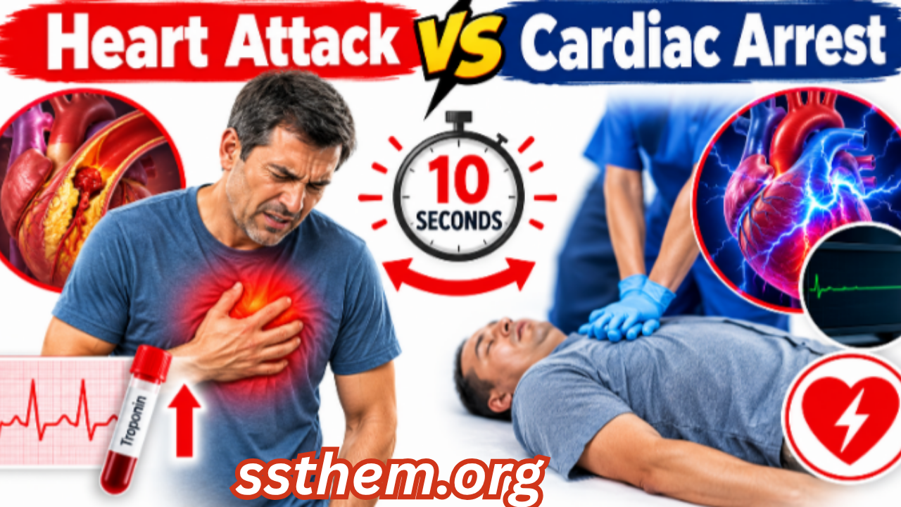
Heart Attack vs Cardiac Arrest 4 Minutes to Act, 10 Minutes to Save a Heart
Heart Attack vs Cardiac Arrest The 10-Second Difference That Can Save a Life — And the Tests That Confirm It
A patient arrives at the emergency department with a symptom that appears routine: a burning discomfort in the upper abdomen. The first ECG looks unremarkable. The initial troponin returns within the normal range. It would be easy to conclude that this is indigestion. Yet the real diagnostic question has only just begun: what evidence can separate a myocardial infarction from the conditions that look remarkably similar? The answer lies not in a single test, but in a disciplined diagnostic process that unfolds over minutes and hours. A heart attack is a circulation problem — a coronary artery becomes blocked, and the heart muscle downstream begins to starve of oxygen. A cardiac arrest is an electrical problem — the heart abruptly stops beating effectively. A heart attack can lead to a cardiac arrest, but they are not the same event. Recognising the difference, and diagnosing a heart attack accurately, is what saves heart muscle and sometimes saves a life.
This is a hypothetical educational case, but it reflects a daily reality in clinical practice. Acute Myocardial Infarction — commonly called a heart attack — remains a leading cause of death worldwide, yet its diagnosis is rarely as simple as a single normal test. The consequences of both overdiagnosis and underdiagnosis are substantial: unnecessary invasive procedures on one hand, and preventable myocardial damage, heart failure, and sudden cardiac death on the other. Accurate diagnosis requires a systematic approach that integrates clinical history, electrocardiography, cardiac biomarkers, imaging, and careful clinical reasoning.
A normal troponin does not always mean the disease is absent.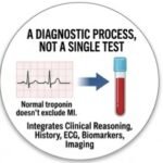 Diagnosis is a process, not a single number on a laboratory report.
This article provides a comprehensive, evidence-based guide to Heart Attack vs Cardiac Arrest, written for medical students, residents, healthcare professionals, and informed readers who want to understand how clinicians separate true myocardial infarction from the conditions that mimic it. If you want to understand why laboratory values can sometimes be misleading, our detailed guide to medical diagnosis explained in 15 powerful steps explains how clinicians interpret these results in context.
Key Diagnostic Facts About Heart Attack (Acute Myocardial Infarction)
The following table summarizes the most clinically important facts about heart attack diagnosis. This is not a substitute for full clinical evaluation, but it provides a rapid reference for healthcare professionals and students.
| Diagnostic Feature | Key Fact |
|---|---|
| Disease | Acute Myocardial Infarction (MI), commonly called a heart attack |
| Main clinical suspicion | Acute chest pain or discomfort, often pressure-like, lasting more than a few minutes, or atypical symptoms in higher-risk groups |
| Most important symptoms | Chest pain or pressure; radiation to arm, jaw, or back; dyspnoea; diaphoresis; nausea; atypical presentations are common in women, older adults, and people with diabetes |
| Important risk factors | Coronary artery disease, hypertension, diabetes, smoking, dyslipidaemia, family history of premature cardiovascular disease, obesity, sedentary lifestyle |
| First clinical step | 12-lead electrocardiogram (ECG) within 10 minutes of first medical contact |
| Initial investigations | ECG; serial high-sensitivity cardiac troponin (hs-cTn) measurements; focused history and physical examination |
| Important laboratory tests | High-sensitivity cardiac troponin (T or I); complete blood count; electrolytes; renal function; glucose; lipid profile |
| Important imaging | Echocardiography (for wall motion abnormalities); coronary angiography (for coronary anatomy and occlusion); cardiac MRI in selected cases |
| Confirmatory test | Rise and/or fall of cardiac troponin with at least one value above the 99th percentile upper reference limit (URL), accompanied by evidence of acute myocardial ischaemia |
| Important differential diagnoses | Unstable angina, myocarditis, pericarditis, pulmonary embolism, aortic dissection, gastro-oesophageal reflux disease, musculoskeletal chest pain |
| Major red flags | Ongoing chest pain with haemodynamic instability; ST-segment elevation on ECG; new left bundle branch block with ischaemic symptoms; ventricular arrhythmias |
| Specialist referral | Immediate cardiology consultation for suspected STEMI; urgent cardiology evaluation for NSTEMI |
| Diagnostic challenge | A single normal troponin does not exclude MI; ECG can be nondiagnostic or normal; atypical presentations are common |
This table is a snapshot. Every parameter in it will be expanded in the dedicated sections below.
Step 1 — When Should Doctors Suspect a Heart Attack?
Typical Presentation: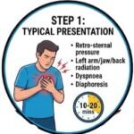 The initial suspicion of acute myocardial infarction rests on recognizing a constellation of clinical features. The classic presentation is retrosternal chest pain or discomfort described as pressure, tightness, squeezing, or heaviness. This discomfort may radiate to the left arm, both arms, the jaw, neck, shoulder, or upper back. It is often accompanied by dyspnoea, diaphoresis, nausea, or a sense of impending doom.
Atypical Presentation: 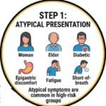 The classic presentation is far from universal. Women, older adults, and individuals with diabetes are more likely to present with atypical symptoms: unexplained fatigue, shortness of breath without chest pain, epigastric discomfort that resembles indigestion, or isolated jaw or back pain. These anginal equivalents should be evaluated with the same diagnostic rigour as typical chest pain.
Duration and Course: Anginal discomfort that lasts more than 10–20 minutes, persists at rest, or awakens the patient from sleep is concerning. Symptoms that are new, occur at lower levels of exertion than before, or are increasing in frequency or severity also raise suspicion. The features that increase clinical suspicion include haemodynamic instability, signs of heart failure, arrhythmias, or a history of established coronary artery disease.
Nonspecific Findings: Many findings associated with myocardial infarction are nonspecific. A patient may describe the discomfort as “indigestion” or “muscle strain.” The presence of risk factors alone does not make the diagnosis, and their absence does not exclude it. How doctors distinguish a heart attack from acid reflux is a common clinical challenge: the two can produce nearly identical symptoms, and a therapeutic trial of antacids does not reliably differentiate them. The diagnostic process, not a single symptom, provides the answer. If you want to understand why some people with significant coronary artery disease never experience classic chest pain, our detailed guide to the 7 silent signs of coronary artery disease explains how the disease can progress quietly for years.
Step 2 — Medical History
A focused but thorough history is the foundation of cardiac diagnosis. The clinician should elicit the following:
- Presenting complaint: Nature, location, radiation, quality, and severity of the symptom.
- Onset: Sudden versus gradual; whether it occurred at rest, during exertion, or during emotional stress.
- Duration and progression: How long the symptom has lasted and whether it is constant or intermittent.
- Associated symptoms: Dyspnoea, diaphoresis, nausea, vomiting, palpitations, syncope, or presyncope.
- Previous episodes: History of angina, prior myocardial infarction, or coronary revascularisation.
- Cardiovascular risk factors: Hypertension, diabetes, dyslipidaemia, smoking, family history of premature coronary artery disease.
- Medications: Current antiplatelet agents, anticoagulants, antihypertensives, lipid-lowering therapy, and any recent changes.
- Social history: Smoking, alcohol use, recreational drug use (particularly cocaine or methamphetamine).
- Family history: Premature cardiovascular disease in first-degree relatives.
The history should also explore features that suggest alternative diagnoses, such as pleuritic pain (pulmonary embolism or pericarditis), tearing pain radiating to the back (aortic dissection), or positional pain relieved by leaning forward (pericarditis). If you want to understand how blood sugar clues can signal early metabolic disease — often coexisting with heart attack risk — our detailed guide to diabetes blood sugar clues and early signs explains the diagnostic overlap in a clear, practical way.
Physical Examination
Vital Signs and General Appearance: The physical examination in suspected myocardial infarction serves several purposes: to assess haemodynamic stability, to identify complications, and to uncover findings that point toward alternative diagnoses. Tachycardia, bradycardia, hypotension, or hypertension may be present. Oxygen saturation should be measured. General appearance may reveal anxiety, diaphoresis, pallor, or signs of shock.
For online earning ideas, freelancing, affiliate marketing, YouTube, Facebook monetization, and AI tips, visit ssthem.xyz for online earning and WhatsApp group links
Cardiovascular Examination: Assess jugular venous pressure (elevated in right ventricular infarction or heart failure), heart sounds (a new murmur may suggest ischaemic mitral regurgitation or ventricular septal defect), and peripheral pulses (asymmetric pulses raise suspicion for aortic dissection). An S4 gallop may suggest left ventricular hypertrophy. A displaced apical impulse may indicate ventricular enlargement.
Respiratory Examination: May reveal crackles consistent with pulmonary oedema. Findings suggesting complications include a new systolic murmur, signs of cardiogenic shock (hypotension, cool extremities, altered mental status), or arrhythmias.
Findings Suggesting Alternative Diagnoses: Pleuritic rub (pericarditis), unilateral leg swelling (deep vein thrombosis with pulmonary embolism), or reproducible chest wall tenderness (musculoskeletal pain).
Red Flags That Should Not Be Ignored
The following table summarizes clinically meaningful warning signs that require urgent attention. These are not exaggerated risks but genuine indicators of potentially serious underlying pathology.
| Red Flag | Why It Matters | Possible Concern | Appropriate Action |
|---|---|---|---|
| ST-segment elevation on ECG | Indicates acute coronary occlusion; time-critical | STEMI | Immediate reperfusion strategy; activate catheterization laboratory |
| Ongoing chest pain with haemodynamic instability | Suggests large infarct or mechanical complication | Cardiogenic shock, ventricular septal rupture, papillary muscle rupture | Urgent cardiology and critical care involvement |
| New left bundle branch block with ischaemic symptoms | May mask STEMI; associated with high mortality | Acute coronary occlusion | Treat as STEMI equivalent until proven otherwise |
| Ventricular tachycardia or fibrillation | Life-threatening arrhythmia | Electrical instability from ischaemia | Immediate defibrillation and advanced cardiac life support |
| Signs of heart failure (pulmonary oedema, elevated JVP) | Indicates extensive myocardial damage | Large anterior MI, right ventricular infarction | Urgent echocardiography and cardiology consultation |
| Syncope or presyncope | May reflect arrhythmia or severe hypotension | High-risk ACS | Continuous cardiac monitoring and urgent evaluation |
Differential Diagnosis
The following table outlines the most important conditions that can resemble a heart attack, along with their distinguishing features and key investigations.
| Differential Diagnosis | Similar Features | Distinguishing Features | Key Investigation |
|---|---|---|---|
| Unstable angina | Chest pain at rest or with minimal exertion; similar quality | No troponin elevation; ECG may show transient changes | Serial troponin; ECG monitoring |
| Myocarditis | Chest pain, troponin elevation, ECG changes | Younger patients; viral prodrome; diffuse ST changes; no coronary occlusion | Cardiac MRI; coronary angiography |
| Pericarditis | Chest pain, ECG changes | Pleuritic pain; positional; diffuse ST elevation with PR depression; friction rub | Echocardiography; ECG pattern |
| Pulmonary embolism | Dyspnoea, chest pain, tachycardia | Pleuritic pain; risk factors for VTE; hypoxaemia; right heart strain | CT pulmonary angiography; D-dimer |
| Aortic dissection | Severe chest pain | Tearing quality; radiation to back; pulse differential; mediastinal widening | CT angiography |
| Gastro-oesophageal reflux disease | Burning chest or epigastric pain | Relation to meals; relief with antacids; no troponin elevation | Clinical response; endoscopy if indicated |
| Musculoskeletal chest pain | Chest wall pain | Reproducible on palpation; positional; no troponin elevation | Clinical examination |
| Anxiety or panic attack | Chest tightness, dyspnoea, palpitations | Younger patients; no risk factors; symptoms reproducible with hyperventilation | Diagnosis of exclusion; troponin and ECG |
The clinical reasoning behind these distinctions is crucial. A normal troponin does not exclude unstable angina, which is a clinical diagnosis based on symptoms and ECG changes. Myocarditis and myocardial infarction can be nearly indistinguishable at presentation, and cardiac MRI is often the key to differentiation. How doctors distinguish a heart attack from acid reflux relies on the overall clinical picture, not on a single symptom or a response to antacids.
Diagnostic Tests
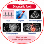
1. 12-Lead Resting Electrocardiogram (ECG): Ordered as the first-line diagnostic tool in suspected myocardial infarction. It is rapid, noninvasive, and can identify ST-segment elevation myocardial infarction (STEMI) within minutes. It records the electrical activity of the heart. In myocardial infarction, it can show ST-segment elevation, ST-segment depression, T-wave inversion, or new Q waves, depending on the stage and type of infarction. A 12-lead ECG should be obtained within 10 minutes of arrival in the emergency department. ECG changes in heart attack include new ST-segment elevation in two or more contiguous leads (≥1 mm in all leads other than V2–V3, where higher thresholds apply for men under 40), new ST-segment depression, T-wave inversion, or new pathological Q waves. A completely normal ECG does not exclude myocardial infarction, particularly in NSTEMI. Can an ECG be normal during a heart attack? Yes—a normal or nondiagnostic ECG is possible, especially in non-ST-elevation presentations. The ECG can be confounded by pre-existing bundle branch block, left ventricular hypertrophy, pacemaker rhythms, or early repolarization patterns. ST elevation can occur in pericarditis, early repolarization, or ventricular aneurysm. NSTEMI and posterior infarction may not produce classic ST elevation. ECG findings must be interpreted alongside troponin results and clinical presentation.
2. High-Sensitivity Cardiac Troponin (hs-cTn): Cardiac troponin is the preferred biomarker for myocardial injury. It is highly specific to cardiac muscle and is released when myocardial cells are damaged. It measures the concentration of troponin T or I in the blood. High-sensitivity assays can detect very low concentrations, enabling earlier diagnosis. It is useful in all patients with suspected acute coronary syndrome. Serial measurements are essential to detect a rise and/or fall pattern. A heart attack blood test result showing a rise and/or fall of troponin with at least one value above the 99th percentile upper reference limit (URL) supports the diagnosis when accompanied by evidence of ischaemia. The troponin levels for heart attack are assay-specific; the 99th percentile URL is the diagnostic threshold, and sex-specific thresholds are recommended. A troponin value below the assay’s limit of detection on a high-sensitivity assay, particularly when measured at least 3 hours after symptom onset, makes myocardial infarction very unlikely. Troponin elevation indicates myocardial injury, not necessarily infarction. Many conditions—myocarditis, pulmonary embolism, heart failure, renal failure, sepsis—can elevate troponin. Elevated troponin may reflect non-ischaemic myocardial injury rather than infarction. A single value is rarely sufficient; how to interpret troponin test results for a heart attack requires understanding that serial testing improves sensitivity.
3. Blood Tests Beyond Troponin: A standard laboratory panel in suspected myocardial infarction includes full blood count (leukocytosis is nonspecific but common), electrolytes (potassium and magnesium abnormalities can cause ECG changes), renal function (creatinine and estimated glomerular filtration rate, important before contrast imaging), glucose and lipid profile (cardiovascular risk assessment), and inflammatory markers such as C-reactive protein (CRP) which may be elevated but are nonspecific. These tests do not diagnose myocardial infarction directly but inform risk assessment, identify exacerbating conditions, and guide safe investigation.
4. Echocardiography: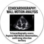 Ordered to assess regional wall motion abnormalities, which can support the diagnosis of myocardial infarction and identify complications. It visualizes cardiac structure, function, and wall motion. New regional wall motion abnormalities in a coronary territory are suggestive of infarction. It is useful when the diagnosis is uncertain, when complications are suspected, or when ECG and troponin results are discordant. A new wall motion abnormality in a vascular territory supports the diagnosis. Normal wall motion does not exclude small infarcts or NSTEMI. Image quality can be suboptimal; findings are operator-dependent.
5. Coronary Angiography: 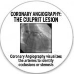 Ordered to visualize the coronary arteries and identify occlusions or stenoses. It assesses coronary anatomy and can identify the culprit lesion. It is the definitive test for patients with STEMI and is used urgently in NSTEMI based on risk stratification. Identification of a coronary thrombus or occlusive lesion confirms the diagnosis. Normal coronary arteries suggest MINOCA (myocardial infarction with non-obstructive coronary arteries) or an alternative diagnosis. It is invasive and carries small risks of complications.
Laboratory Tests Table
| Test | Purpose | Possible Result | Interpretation | Limitations |
|---|---|---|---|---|
| hs-cTn | Detect myocardial injury | Rise/fall above 99th percentile | Supports MI in ischaemic context | Elevated in non-ischaemic conditions |
| Full blood count | Assess for leukocytosis | Leukocytosis common in acute MI | Nonspecific; supports inflammatory response | Nonspecific |
| Renal function | Assess kidney function | Reduced eGFR indicates chronic kidney disease | Essential baseline before contrast imaging | eGFR equations vary by population |
| Glucose and HbA1c | Screen for diabetes | Elevated glucose or HbA1c indicates diabetes or prediabetes | Comorbidity affecting risk and treatment | HbA1c affected by haemoglobin variants |
| Lipid profile | Assess cardiovascular risk | Elevated LDL, low HDL increase risk | Guides statin therapy decisions | Fasting status may affect results |
| Electrolytes | Assess for abnormalities | Potassium and magnesium abnormalities can cause ECG changes | Guides safe investigation | Affected by medications, hydration |
Imaging Studies
Chest X-ray: Not used to diagnose myocardial infarction but may reveal cardiomegaly, pulmonary oedema, or alternative causes of chest pain. It provides a baseline assessment of cardiac silhouette and pulmonary vasculature.
Echocardiography: Assesses cardiac structure and function, identifying complications and alternative diagnoses. It is useful in patients with heart failure symptoms, murmurs, or abnormal ECG findings. Regional wall motion abnormalities in a coronary territory distribution may support the diagnosis. Normal wall motion and function do not exclude myocardial infarction.
Coronary Angiography: Remains the reference standard for anatomical assessment and guides revascularisation. It provides direct visualisation of the coronary lumen and allows measurement of FFR/iFR for functional assessment. In STEMI, immediate coronary angiography is the standard of care.
Cardiac MRI: Used in selected cases to differentiate myocardial infarction from myocarditis, to assess myocardial viability, and to identify complications. Late gadolinium enhancement in a coronary territory pattern supports the diagnosis of infarction. It is the gold standard for detecting prior myocardial scar.
If you want to understand how hypertension diagnosis can overlap with heart attack risk assessment, our detailed guide to hypertension diagnosis explained with BP readings and home monitoring explains the diagnostic criteria in a clear, practical way.
Specialized and Confirmatory Testing
Invasive coronary functional testing may be used to assess the haemodynamic significance of intermediate lesions. This includes fractional flow reserve (FFR) and instantaneous wave-free ratio (iFR).
Intravascular ultrasound (IVUS) and optical coherence tomography (OCT) characterise plaque and optimise PCI. They are used in complex lesions, left main disease, or when angiography is ambiguous.
Cardiac MRI is the gold standard for detecting prior myocardial scar and can differentiate acute myocardial infarction from myocarditis. It is useful when the diagnosis remains uncertain after initial testing.
Diagnostic Criteria
The diagnosis of acute myocardial infarction is defined by the Fourth Universal Definition of Myocardial Infarction (2018), developed by the European Society of Cardiology (ESC), American College of Cardiology (ACC), American Heart Association (AHA), and World Heart Federation (WHF). The Fifth Universal Definition was published in 2026 and builds upon the same foundational principles.
The diagnostic criteria for type 1 (spontaneous) myocardial infarction require: (1) Detection of a rise and/or fall of cardiac troponin values with at least one value above the 99th percentile upper reference limit (URL); and (2) At least one of the following: symptoms of myocardial ischaemia; new ischaemic ECG changes; development of pathological Q waves; imaging evidence of new loss of viable myocardium or new regional wall motion abnormality in a pattern consistent with an ischaemic etiology; identification of a coronary thrombus by angiography or autopsy.
It is critical to understand that an elevated troponin alone is not sufficient to diagnose myocardial infarction. Myocardial injury—elevated troponin without ischaemic evidence—is a separate condition. Type 2 myocardial infarction occurs when there is an imbalance between oxygen supply and demand, not due to acute coronary thrombosis. The 99th percentile URL is the diagnostic threshold, but it is assay-specific and should be sex-specific. Reference values vary between laboratories and assays, and age-related differences are important: approximately 1 in 3 patients over 75 with possible myocardial infarction have an elevated troponin compared to 1 in 20 patients under 50.
Diagnostic Algorithm
| Step | Action | Key Consideration |
|---|---|---|
| 1. Symptom recognition | Chest pain, dyspnoea, atypical symptoms in high-risk groups | Assess symptom quality, duration, and provocation |
| 2. 12-lead ECG within 10 minutes | Identify ST elevation or other ischaemic changes | STEMI requires immediate reperfusion |
| 3. Focused history and examination | Assess risk factors, symptom characteristics, haemodynamic status | Determine pretest probability |
| 4. Initial troponin | Draw at presentation; repeat at 1–3 hours per protocol | Serial testing essential; single value may be normal |
| 5. ECG interpretation | STEMI vs NSTEMI vs nondiagnostic | New LBBB with symptoms is STEMI equivalent |
| 6. Differential diagnosis | Exclude pericarditis, myocarditis, PE, dissection, GI causes | Clinical context guides testing |
| 7. Echocardiography | Assess wall motion, complications, alternative diagnoses | Useful when diagnosis uncertain |
| 8. Coronary angiography | Urgent for STEMI; risk-stratified for NSTEMI | Identify culprit lesion; guide revascularisation |
| 9. Severity assessment | Killip class, haemodynamic status, infarct size | Guides management intensity |
| 10. Specialist referral | Cardiology for all confirmed or high-risk cases | Revascularisation vs. medical therapy |
| 11. Follow-up | Secondary prevention, cardiac rehabilitation, risk factor management | Ongoing assessment |
Severity and Staging
Myocardial infarction severity is classified by the extent of myocardial damage and the presence of complications. The Killip classification is commonly used: Killip Class I: No clinical signs of heart failure. Killip Class II: Rales in the lungs, S3 gallop, jugular venous distension. Killip Class III: Acute pulmonary oedema. Killip Class IV: Cardiogenic shock.
Infarct location also influences prognosis: anterior infarcts are generally larger and associated with higher mortality than inferior infarcts. The presence of right ventricular involvement, mechanical complications, or arrhythmias further stratifies risk.
Special Populations
Women: Women are more likely to present with atypical symptoms—jaw or back pain, nausea, fatigue, and dyspnoea—and are more likely to have myocardial infarction with non-obstructive coronary arteries (MINOCA) or spontaneous coronary artery dissection (SCAD). How is a heart attack diagnosed in women? The diagnostic approach is the same, but clinicians must maintain a high index of suspicion when symptoms are not classic.
Young adults: How is a heart attack diagnosed in young adults? The same diagnostic tools are used, but the differential diagnosis is broader. Causes include coronary artery anomalies, SCAD, coronary embolism, and thrombophilia. Traditional risk stratification may underperform in younger patients.
Older adults: Troponin levels are more likely to be elevated due to age-related comorbidities, and symptoms may be atypical or masked by cognitive impairment.
Patients with kidney disease: Troponin levels are often chronically elevated, making interpretation challenging. Serial measurements and clinical context are essential.
Pregnancy: Pregnancy increases cardiac demand and can unmask or exacerbate coronary artery disease. Diagnosis requires careful interpretation of troponin (which may be elevated in preeclampsia) and ECG changes.
False Positive and False Negative Results
False Positive: An elevated troponin may reflect myocardial injury from non-ischaemic causes: myocarditis, pulmonary embolism, heart failure, sepsis, renal failure, or extreme exertion. The clinician must assess whether the elevated troponin is accompanied by evidence of acute myocardial ischaemia. A normal result does not always mean the disease is absent, and an abnormal result does not always confirm the disease.
False Negative: A normal troponin, particularly if drawn very early after symptom onset, does not exclude myocardial infarction. Serial testing is essential. Unstable angina—ischaemia without necrosis—will not elevate troponin but still requires urgent evaluation. Diagnosis is a process, not a single number on a laboratory report.
Clinicians use pretest probability, serial testing, and confirmatory investigations to navigate these uncertainties. The most useful test is not necessarily the most advanced test; it is the one that answers the clinical question in context.
10 Common Diagnostic Mistakes in Heart Attack
- Anchoring on a single normal troponin: A single early troponin does not exclude MI.
- Premature diagnostic closure: Attributing symptoms to a benign cause without completing the evaluation.
- Ignoring atypical presentations: Failing to consider MI in women, older adults, and patients with diabetes.
- Over-relying on the ECG: A normal ECG does not exclude NSTEMI.
- Misunderstanding reference ranges: Using a uniform troponin threshold rather than assay-specific and sex-specific values.
- Ignoring pretest probability: Ordering troponin indiscriminately without clinical context.
- Failing to consider differential diagnoses: Missing myocarditis, pulmonary embolism, or aortic dissection.
- Inappropriate test selection: Using non-high-sensitivity troponin when hs-cTn is available.
- Misinterpreting false-positive results: Treating troponin elevation as synonymous with MI.
- Failing to reassess an evolving patient: Symptoms and ECG findings can change rapidly.
Hypothetical Clinical Cases
These are hypothetical educational cases and do not represent real patients.
Case 1: The Atypical Presentation
Presentation: A 58-year-old woman with a history of hypertension and type 2 diabetes presents with nausea, diaphoresis, and jaw pain that began two hours ago. She denies chest pain.
Initial Differential: NSTEMI, unstable angina, gastro-oesophageal reflux, musculoskeletal pain.
History: Symptoms began two hours ago; nausea, diaphoresis, and jaw pain. No chest pain. Risk factors: hypertension, type 2 diabetes.
Examination: Heart rate 92 bpm, blood pressure 145/88 mmHg, diaphoretic.
Investigations: ECG shows nonspecific ST-T changes. Initial troponin mildly elevated. Serial troponin shows a rise. Echocardiography reveals a regional wall motion abnormality. Coronary angiography identifies a critical stenosis in the left circumflex artery.
Clinical Reasoning: The combination of risk factors, atypical symptoms, and troponin elevation raises suspicion for NSTEMI.
Diagnosis: NSTEMI due to atherosclerotic coronary artery disease.
Key Diagnostic Lesson: Atypical symptoms in women with risk factors warrant a full cardiac evaluation.
Case 2: The Reassuring ECG
Presentation: A 45-year-old man with a history of smoking presents with severe epigastric pain radiating to the back. ECG is normal. Initial troponin is normal. He is initially thought to have gastritis.
Initial Differential: Gastritis, GERD, musculoskeletal pain, NSTEMI.
History: Severe epigastric pain radiating to the back. Smoking history.
Examination: Heart rate 88 bpm, blood pressure 138/86 mmHg, epigastric tenderness.
Investigations: Initial ECG normal. Initial troponin normal. Two hours later, troponin has risen significantly, and repeat ECG shows new ST depression in the lateral leads. Coronary angiography showed a severe stenosis in the right coronary artery.
Clinical Reasoning: The initial normal ECG and troponin were falsely reassuring. Serial testing revealed evolving myocardial injury.
Diagnosis: NSTEMI.
Key Diagnostic Lesson: Serial troponin testing is essential; a single normal result does not exclude MI.
Case 3: The Silent Infarction
Presentation: A 72-year-old man with diabetes and no prior cardiac history presents for a routine check-up. He mentions occasional fatigue but no chest pain.
Initial Differential: Prior silent MI, heart failure, anaemia.
History: Diabetes, no prior cardiac history. Occasional fatigue.
Examination: Blood pressure 138/84 mmHg, heart rate 72 bpm. No murmurs.
Investigations: ECG shows pathological Q waves in the inferior leads. Echocardiography reveals inferior wall hypokinesis. Cardiac MRI confirms scar tissue.
Clinical Reasoning: How is a silent heart attack diagnosed? Unrecognized myocardial infarction is identified through ECG findings (Q waves) or imaging evidence of prior infarction in a patient without a known history.
Diagnosis: Prior silent myocardial infarction.
Key Diagnostic Lesson: Silent myocardial infarction is common in patients with diabetes and should be considered when ECG or imaging findings suggest prior injury.
Patient-Friendly Diagnosis Guide
What Should a Patient Expect During Diagnosis?
When you arrive at the emergency department with symptoms that could be a heart attack, the evaluation begins immediately. You will be connected to a cardiac monitor, and a 12-lead ECG will be performed within minutes. Blood will be drawn for troponin testing, and this may be repeated after a few hours.
You may undergo an echocardiogram, which uses sound waves to create a picture of your heart. If a heart attack is confirmed, you may be taken for a coronary angiogram—a procedure that uses dye and X-rays to visualize the arteries supplying your heart. This can also be used to open a blocked artery.
Waiting for results can be stressful. The medical team is not withholding information; they are gathering the evidence needed to make an accurate diagnosis. Additional tests may be needed if the initial results are unclear. Follow-up appointments are important to manage risk factors and prevent future events.
When to Seek Urgent Medical Care
Seek emergency medical care immediately if you experience:
- Chest pain or discomfort that lasts more than a few minutes, or that goes away and comes back
- Pain or discomfort in one or both arms, the jaw, neck, or back
- Shortness of breath, with or without chest discomfort
- Nausea, vomiting, or diaphoresis
- Lightheadedness or syncope
Do not attempt to drive yourself to the hospital. Call emergency services. Do not take nitroglycerin or other medications without medical advice if you are unsure of your diagnosis.
1. A 62-year-old man presents with central chest pressure and diaphoresis. His ECG shows ST elevation in leads II, III, and aVF. What is the most likely diagnosis?
A. Anterior STEMI
B. Inferior STEMI
C. Pericarditis
D. Pulmonary embolism
Correct Answer: B
Explanation: ST elevation in leads II, III, and aVF indicates an inferior wall STEMI, typically due to right coronary artery occlusion.
2. Which troponin threshold is used to define myocardial injury?
A. 50th percentile
B. 75th percentile
C. 95th percentile
D. 99th percentile upper reference limit
Correct Answer: D
Explanation: The 99th percentile URL is the standardized threshold for myocardial injury and infarction.
3. A patient’s troponin is elevated but ECG is normal and symptoms are absent. What is the most appropriate interpretation?
A. Definite myocardial infarction
B. Myocardial injury
C. Normal variant
D. Unstable angina
Correct Answer: B
Explanation: Elevated troponin without ischaemic evidence defines myocardial injury, not infarction.
4. Which finding on ECG suggests pericarditis rather than STEMI?
A. ST elevation in contiguous leads
B. PR depression with diffuse ST elevation
C. Pathological Q waves
D. New left bundle branch block
Correct Answer: B
Explanation: PR depression with diffuse ST elevation is characteristic of pericarditis.
5. A 28-year-old woman presents with chest pain and troponin elevation. Coronary angiography is normal. What diagnosis should be considered?
A. STEMI
B. MINOCA
C. Stable angina
D. Aortic dissection
Correct Answer: B
Explanation: MINOCA (myocardial infarction with non-obstructive coronary arteries) should be considered when troponin is elevated but coronary arteries are normal.
6. What is the recommended time window for obtaining a 12-lead ECG in suspected MI?
A. Within 30 minutes
B. Within 10 minutes
C. Within 1 hour
D. Within 2 hours
Correct Answer: B
Explanation: Current guidelines recommend ECG within 10 minutes of first medical contact.
7. Which symptom is most common in women presenting with MI?
A. Isolated jaw pain
B. Chest pain
C. Nausea
D. Fatigue
Correct Answer: B
Explanation: Although women may have atypical symptoms, chest pain remains the most common presentation.
8. A patient’s troponin is normal at 1 hour after symptom onset. What is the next step?
A. Discharge home
B. Repeat troponin at 3 hours
C. Immediate angiography
D. Start thrombolysis
Correct Answer: B
Explanation: Serial troponin testing is essential because a single early value may be falsely negative.
9. Which condition can cause false-positive troponin elevation?
A. Stable angina
B. Pulmonary embolism
C. Musculoskeletal pain
D. GERD
Correct Answer: B
Explanation: Pulmonary embolism can cause myocardial injury and troponin elevation.
10. What ECG finding is diagnostic of STEMI?
A. T-wave inversion in V4–V6
B. ST depression in II, III, aVF
C. ST elevation ≥1 mm in two contiguous leads
D. Q waves in V1–V2
Correct Answer: C
Explanation: New ST elevation ≥1 mm in two contiguous leads (with higher thresholds for V2–V3 in men under 40) is diagnostic of STEMI.
11. A 70-year-old man with diabetes presents with fatigue and dyspnoea. ECG shows Q waves in V1–V4. What is the most likely diagnosis?
A. Acute anterior STEMI
B. Prior anterior myocardial infarction
C. Pericarditis
D. Pulmonary embolism
Correct Answer: B
Explanation: Pathological Q waves indicate prior infarction; acute injury would show ST changes.
12. Which test is the gold standard for detecting prior myocardial scar?
A. ECG
B. Echocardiography
C. Cardiac MRI
D. Chest X-ray
Correct Answer: C
Explanation: Cardiac MRI with late gadolinium enhancement is the gold standard for visualizing myocardial scar.
13. A patient presents with tearing chest pain radiating to the back. What diagnosis must be excluded?
A. STEMI
B. Aortic dissection
C. Pericarditis
D. GERD
Correct Answer: B
Explanation: Tearing pain radiating to the back is classic for aortic dissection.
14. Which biomarker is preferred for MI diagnosis?
A. CK-MB
B. Myoglobin
C. Cardiac troponin
D. LDH
Correct Answer: C
Explanation: Cardiac troponin is the preferred biomarker due to its high sensitivity and specificity.
15. What is the first step in managing a patient with suspected STEMI?
A. Wait for troponin results
B. Obtain ECG and activate catheterization laboratory
C. Start anticoagulation
D. Order echocardiography
Correct Answer: B
Explanation: Immediate ECG and reperfusion strategy are critical in STEMI.
16. Which population is most likely to have elevated troponin at baseline?
A. Young athletes
B. Patients with renal failure
C. Healthy children
D. Pregnant women in first trimester
Correct Answer: B
Explanation: Renal failure is associated with chronically elevated troponin levels.
17. A patient with normal coronary arteries on angiography and elevated troponin may have:
A. Stable angina
B. MINOCA
C. GERD
D. Costochondritis
Correct Answer: B
Explanation: MINOCA is diagnosed when troponin is elevated but coronary arteries are normal.
18. Which ECG change is a STEMI equivalent?
A. T-wave inversion
B. New left bundle branch block with ischaemic symptoms
C. First-degree AV block
D. Sinus tachycardia
Correct Answer: B
Explanation: New LBBB with ischaemic symptoms is treated as a STEMI equivalent.
19. What is the most important limitation of troponin testing?
A. It is invasive
B. It is not specific for ischaemia
C. It requires fasting
D. It is only available in hospitals
Correct Answer: B
Explanation: Troponin elevation indicates myocardial injury, not necessarily ischaemic infarction.
20. A patient with suspected MI has a normal ECG and normal troponin at 3 hours. What is the most appropriate next step?
A. Discharge with reassurance
B. Consider alternative diagnoses and reassess
C. Immediate angiography
D. Start thrombolysis
Correct Answer: B
Explanation: Alternative diagnoses should be considered, and the patient should be reassessed if symptoms persist.
21. Which symptom is most suggestive of myocardial ischaemia in a patient with diabetes?
A. Classic chest pain
B. Exertional dyspnoea
C. Sharp pleuritic pain
D. Positional pain
Correct Answer: B
Explanation: Patients with diabetes often present with dyspnoea rather than classic chest pain.
22. What is the role of echocardiography in suspected MI?
A. Confirm coronary occlusion
B. Assess wall motion abnormalities and complications
C. Replace troponin testing
D. Diagnose pulmonary embolism
Correct Answer: B
Explanation: Echocardiography assesses regional wall motion and complications but does not visualize coronary arteries.
23. Which condition can mimic MI with ST elevation?
A. Early repolarization
B. Atrial fibrillation
C. Sinus bradycardia
D. First-degree AV block
Correct Answer: A
Explanation: Early repolarization can cause benign ST elevation that mimics STEMI.
24. What is the definition of type 2 myocardial infarction?
A. MI due to coronary thrombosis
B. MI due to supply-demand mismatch
C. MI due to coronary embolism
D. MI due to spontaneous coronary dissection
Correct Answer: B
Explanation: Type 2 MI is caused by an imbalance between myocardial oxygen supply and demand, not acute coronary thrombosis.
25. A patient with known coronary artery disease presents with chest pain at rest. ECG is normal, and troponin is negative. What is the most likely diagnosis?
A. Unstable angina
B. STEMI
C. Pericarditis
D. Aortic dissection
Correct Answer: A
Explanation: Unstable angina is characterized by ischaemic symptoms without troponin elevation or diagnostic ECG changes.
Question. How is a heart attack diagnosed?
Answer : A heart attack is diagnosed through a combination of clinical history, ECG findings, and cardiac troponin levels. The diagnosis requires a rise and/or fall of troponin with at least one value above the 99th percentile URL, accompanied by evidence of acute myocardial ischaemia.
Question. What is the first test for a heart attack?
Answer : The first test is a 12-lead ECG, which should be performed within 10 minutes of first medical contact.
Question. What blood test confirms a heart attack?
Answer : High-sensitivity cardiac troponin (T or I) is the preferred blood test. Serial measurements are used to detect a rise and/or fall pattern.
Question. Can a heart attack be diagnosed without a troponin test?
Answer : In some cases, STEMI can be diagnosed based on ECG findings alone. However, troponin is essential for NSTEMI diagnosis and for distinguishing myocardial injury from infarction.
Question. What does a positive troponin mean?
Answer : A positive troponin indicates myocardial injury. It does not automatically confirm myocardial infarction; the result must be interpreted with clinical and ECG evidence of ischaemia.
Question. Can troponin tests be falsely negative?
Answer : Yes. A single early troponin may be normal. Serial testing and clinical assessment are essential.
Question. Can troponin tests be falsely positive?
Answer : Troponin elevation can occur in many conditions other than MI, including myocarditis, pulmonary embolism, heart failure, and renal failure.
Question. When is imaging needed?
Answer : Echocardiography is used when the diagnosis is uncertain or complications are suspected. Coronary angiography is used for confirmed or high-risk cases.
Question. When is a specialist consulted?
Answer : Cardiology consultation is standard for all confirmed or high-risk cases of myocardial infarction.
Question. How long does it take to diagnose a heart attack?
Answer : The diagnosis can be confirmed within 1–3 hours using serial troponin testing and ECG. STEMI can be diagnosed within minutes of ECG acquisition.
Question. How is a silent heart attack diagnosed?
Answer : Silent myocardial infarction is diagnosed through ECG findings (Q waves) or imaging evidence of prior infarction in a patient without a known history. Cardiac MRI is the gold standard for confirming scar.
Question. How is a heart attack diagnosed in women?
Answer : The diagnostic approach is the same, but clinicians must maintain a high index of suspicion for atypical symptoms, including jaw or back pain, nausea, and fatigue.
Question. What is the difference between a heart attack and angina diagnosis?
Answer : Unstable angina causes ischaemic symptoms without troponin elevation. Myocardial infarction involves myocardial necrosis with troponin elevation.
Question. How do doctors distinguish a heart attack from acid reflux?
Answer : The distinction relies on the overall clinical picture, including risk factors, symptom characteristics, ECG findings, and troponin results. A response to antacids does not reliably differentiate the two.
Question. When should urgent medical care be sought?
Answer : Seek emergency care for chest pain lasting more than a few minutes, pain radiating to the arm or jaw, shortness of breath, diaphoresis, or syncope.
Latest Evidence & References
- Thygesen K, Alpert JS, Jaffe AS, et al. Fourth Universal Definition of Myocardial Infarction (2018). European Heart Journal. 2018. Published simultaneously in Circulation and Journal of the American College of Cardiology. DOI: 10.1093/eurheartj/ehy462.
- Thygesen K, Alpert JS, Jaffe AS, et al. Fifth Universal Definition of Myocardial Infarction (2026). Circulation. 2026. DOI: 10.1161/CIR.0000000000001477.
- American College of Cardiology. Fourth Universal Definition of MI Released. ACC Latest in Cardiology. August 2018.
- Centers for Disease Control and Prevention. About Coronary Artery Disease (CAD). CDC Heart Disease. Updated February 2025.
- Centers for Disease Control and Prevention. Effectiveness of Practices for Improving the Diagnostic Accuracy of Non-ST Elevation Myocardial Infarction in the Emergency Department. CDC Laboratory Medicine Best Practices Systematic Review.
- American Heart Association. Heart Attacks in Younger Women: AHA Calls for Better Diagnosis and Management. JAMA. 2026.
- American Heart Association. Acute Coronary Syndrome in Women. Circulation. 2024.
- Merck Manual Professional Edition. Acute Myocardial Infarction (MI). Updated February 2024.
- National Health Service (UK). Heart Assessment Centre. Imperial College Healthcare NHS Trust.
- National Health Service (UK). Troponin I: Guide for Primary Care. Right Decisions Scotland.
- Rao SV, O’Donoghue ML, Ruel M, et al. 2025 ACC/AHA/ACEP/NAEMSP/SCAI Guideline for the Management of Patients With Acute Coronary Syndromes. Circulation. 2025.
- Gulati M, Levy PD, Mukherjee D, et al. 2021 AHA/ACC/ASE/CHEST/SAEM/SCCT/SCMR Guideline for the Evaluation and Diagnosis of Chest Pain. Circulation. 2021;144(22):e368–e454.
- McDermott M, et al. Cardiac troponin at the point of care in acute and chronic coronary syndromes. Diabetes, Obesity and Metabolism. 2025;27(Suppl 8):47–58.
- Knuuti J, Wijns W, Saraste A, et al. 2019 ESC Guidelines for the diagnosis and management of chronic coronary syndromes. European Heart Journal. 2020;41(3):407–477.
- American College of Cardiology. Top Takeaways From the 2025 ACC/AHA Acute Coronary Syndrome Guideline. 2025.
Medical Information Disclaimer: The information provided in this article is for educational and informational purposes only and is intended for healthcare professionals, medical students, and informed general readers. It does not constitute medical advice, diagnosis, or treatment recommendations. Heart attack is a medical emergency that should be evaluated and managed by a qualified healthcare provider. Treatment decisions, including medication selection, revascularisation, and monitoring, depend on the patient’s individual circumstances, comorbidities, and clinical judgment. Readers should not use this information to self-diagnose, self-treat, or make clinical decisions without appropriate professional consultation. If you have concerns about your heart health, seek evaluation from a qualified healthcare professional. The authors and publishers of this article do not assume any liability for any adverse effects or consequences resulting from the use or misuse of the information provided herein.
For additional health and wellness resources, you may also find evidence-based articles on ssthem.net for health and beauty tips helpful.



3 Comments