10 Powerful Facts About Phase II Metabolism That Every Healthcare Professional Must Know
1. Phase II drug metabolism
Drug metabolism represents one of the most critical determinants of pharmacological response, governing drug efficacy, toxicity, and duration of action. The human body possesses sophisticated enzymatic systems designed to transform lipophilic xenobiotics into more hydrophilic, excretable metabolites. This biotransformation process is broadly classified into Phase I (functionalization) and Phase II (conjugation) reactions.
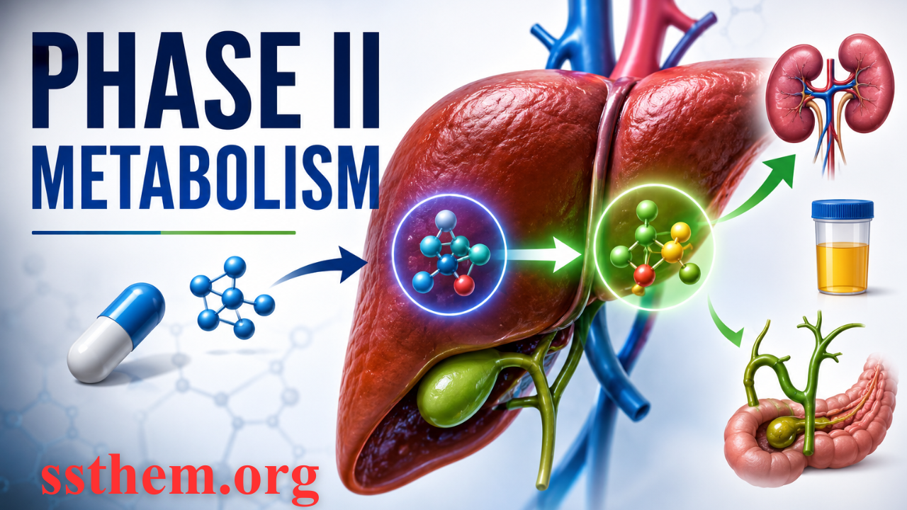
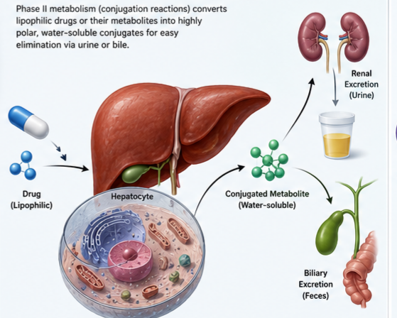
Phase II drug metabolism, often termed conjugation reactions, involves the coupling of a drug or its Phase I metabolite with an endogenous molecule to produce a highly polar, readily excretable conjugate. These reactions are predominantly catalyzed by transferases, enzymes that facilitate the transfer of a conjugating moiety from an activated cofactor to a substrate bearing a suitable functional group.
While Phase I reactions introduce or expose functional groups such as hydroxyl, amino, or carboxyl groups, Phase II reactions capitalize on these chemical handles to attach large, hydrophilic molecules like glucuronic acid, sulfate, or glutathione. The resulting conjugates are typically more water-soluble, have higher molecular weights, and are efficiently eliminated via urine or bile.
This comprehensive guide explores the biochemistry, enzymology, clinical significance, and pharmacogenetic implications of Phase II drug metabolism. Drawing from authoritative sources including peer-reviewed literature, pharmacological textbooks, and regulatory guidance, this article provides healthcare professionals and researchers with an up-to-date understanding of this essential physiological process.
اسلامی و مذہبی معلومات | Religious Knowledge قرآن، حدیث، انبیاء علیہم السلام، اسلامی تاریخ، مختلف مذاہب، تقابلی مطالعہ اور مستند مذہبی معلومات ایک ہی جگہ حاصل کریں۔ اگر آپ اسلام، دیگر مذاہب اور مذہبی تاریخ کے بارے میں تحقیقی اور معلوماتی مضامین پڑھنا چاہتے ہیں تو ضرور وزٹ کریں علم میں اضافہ کریں، تحقیق کریں اور مستند معلومات حاصل کریں۔ssthem.com
2. What is Phase II Metabolism?
2.1 Definition
Phase II metabolism, also referred to as synthetic reactions or conjugation reactions, represents the second phase of drug biotransformation. In these reactions, a drug molecule or its Phase I metabolite is covalently linked to an endogenous molecule—a “conjugating moiety”—resulting in the formation of a conjugate.
The conjugating moiety, typically ranging from 100 to 300 Daltons in molecular weight, is supplied by a high-energy cofactor or co-substrate. These cofactors include:
- UDP-glucuronic acid (for glucuronidation)
- 3′-Phosphoadenosine-5′-phosphosulfate (PAPS) (for sulfation)
- Acetyl-CoA (for acetylation)
- S-Adenosylmethionine (SAM) (for methylation)
- Glutathione (GSH) (for glutathione conjugation)
These conjugation reactions are almost exclusively catalyzed by transferases, with major enzymes including UDP-glucuronosyltransferases (UGTs), sulfotransferases (SULTs), N-acetyltransferases (NATs), glutathione S-transferases (GSTs), and methyltransferases.
2.2 Why Phase II Metabolism is Important
Phase II metabolism serves several critical physiological functions:
1. Detoxification and Elimination
The primary purpose of conjugation is to convert lipophilic compounds into hydrophilic, readily excretable metabolites. This process prevents the accumulation of potentially toxic drugs and xenobiotics in the body.
2. Termination of Pharmacological Activity
For many drugs, Phase II metabolism represents the terminal step in drug clearance, transforming pharmacologically active compounds into inactive conjugates that are eliminated from the body.
3. Prodrug Activation
While most conjugation reactions produce inactive metabolites, certain prodrugs are activated through Phase II pathways. Some glucuronide metabolites retain pharmacological activity or can be cleaved back to the active parent drug at target tissues.
4. Protection Against Reactive Intermediates
Glutathione conjugation, in particular, serves as a crucial defense mechanism against reactive electrophilic metabolites that can cause cellular damage and toxicity.
5. Clinical Drug Interactions
Inhibition or induction of Phase II enzymes can significantly alter drug pharmacokinetics, leading to clinically relevant drug–drug interactions. Understanding these pathways is essential for safe prescribing and therapeutic drug monitoring.
3. Relationship Between Phase I and Phase II Metabolism
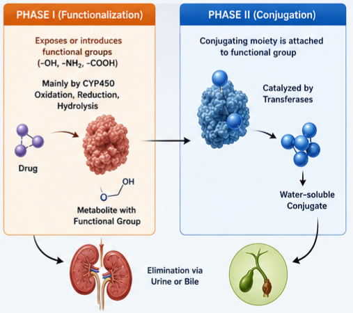 The classic view of drug metabolism posits a sequential process: Phase I reactions introduce a functional group, which then serves as a “chemical anchor” for Phase II conjugation. This model, while useful, has important exceptions:
Sequential Metabolism:
– Phase I (functionalization): CYP450-mediated oxidation, reduction, or hydrolysis introduces or exposes -OH, -NH₂, -COOH groups
– Phase II (conjugation): A conjugating moiety is attached to the newly exposed functional group
– Example: Acetaminophen undergoes CYP450-mediated oxidation to N-acetyl-p-benzoquinone imine (NAPQI), which is then conjugated with glutathione
Direct Phase II Metabolism:
Many drugs containing pre-existing functional groups undergo direct conjugation without prior Phase I metabolism:
– Morphine undergoes direct glucuronidation at the 3- and 6-hydroxyl positions
– Isoniazid is directly acetylated by NAT2
– Salbutamol undergoes direct sulfation
Phase II Followed by Phase I:
In some instances, conjugation may precede Phase I metabolism. For example, certain drugs may undergo glucuronidation followed by oxidation of the glucuronide moiety.
Non-sequential Metabolism:
Phase I metabolites may be eliminated directly without conjugation. Conversely, some Phase II conjugates can undergo further Phase I metabolism.
This complexity underscores that Phase I and Phase II metabolism are not strictly sequential but represent a highly integrated and adaptable detoxification system.
4. Sites of Phase II Metabolism
Phase II drug-metabolizing enzymes are widely distributed throughout the body, reflecting the need for local and systemic detoxification.
4.1 Liver
The liver represents the primary site of Phase II metabolism, expressing high levels of UGTs, SULTs, GSTs, NATs, and methyltransferases. The strategic location of the liver—receiving blood from the gastrointestinal tract via the portal vein—allows it to act as the first line of defense against ingested xenobiotics.
Hepatocellular Compartmentalization:
– Endoplasmic reticulum (luminal side): Glucuronidation (UGTs)
– Cytosol: Sulfation (SULTs), glutathione conjugation (GSTs), acetylation (NATs), methylation (MTs)
4.2 Gastrointestinal Tract
The intestinal wall expresses significant Phase II enzyme activity, particularly UGTs, SULTs, and GSTs. Intestinal metabolism contributes to:
– First-pass metabolism: Reducing oral bioavailability of drugs
– Enterohepatic recirculation: Metabolites formed in the intestine can be reabsorbed or excreted
– Local detoxification: Protecting gut mucosa from dietary toxins and bacterial products
4.3 Kidney
The kidney expresses a range of Phase II enzymes, including UGTs, SULTs, and GSTs. Renal conjugation contributes to:
– Local metabolism: Converting drugs to more polar forms within renal tissue
– Excretion: Conjugates are actively secreted into the proximal tubule for urinary elimination via organic anion transporters (OATs) and multidrug resistance-associated proteins (MRPs)
4.4 Other Tissues
Phase II enzymes are also present in:
– Lungs: Express SULTs and GSTs for detoxification of inhaled xenobiotics
– Skin: Contains UGTs and SULTs for local metabolism
– Brain: Low levels of UGTs, SULTs, and methyltransferases for neurotransmitter metabolism
– Placenta: Provides fetal protection from maternal drugs and toxins
5. Enzymes Involved in Phase II Metabolism
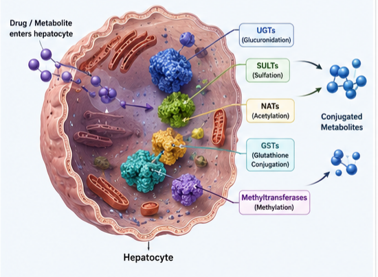
The major Phase II drug-metabolizing enzymes (DMEs) are transferases. Each enzyme family exhibits distinct substrate specificity, tissue distribution, and genetic polymorphism.
Table 1: Major Phase II Drug-Metabolizing Enzymes
| Enzyme Family | Reaction | Cofactor | Subcellular Location | Key Substrates |
|---|---|---|---|---|
| UDP-Glucuronosyltransferases (UGTs) | Glucuronidation | UDP-glucuronic acid | Endoplasmic reticulum | Morphine, bilirubin, acetaminophen, NSAIDs |
| Sulfotransferases (SULTs) | Sulfation | PAPS (3′-phosphoadenosine-5′-phosphosulfate) | Cytosol | Steroids, catecholamines, acetaminophen |
| N-Acetyltransferases (NATs) | Acetylation | Acetyl-CoA | Cytosol | Isoniazid, hydralazine, sulfonamides |
| Glutathione S-Transferases (GSTs) | Glutathione conjugation | Glutathione (GSH) | Cytosol | Reactive electrophiles, carcinogens |
| Methyltransferases (MTs) | Methylation | S-Adenosylmethionine (SAM) | Cytosol/membrane | Catecholamines, thiopurines |
Sources:
5.1 UDP-Glucuronosyltransferases (UGTs)
UGTs represent the most important family of Phase II enzymes, catalyzing the transfer of glucuronic acid from UDP-glucuronic acid to substrates containing hydroxyl, carboxyl, amino, or sulfhydryl groups.
Characteristics:
– Located on the luminal side of the endoplasmic reticulum
– Consist of two main families: UGT1A (sharing exon 1) and UGT2B (multiple exons)
– Over 30 functional UGT isoforms identified in humans
– UGT1A1 is the most clinically significant due to Gilbert’s syndrome and irinotecan toxicity
Clinical Significance:
– Bilirubin conjugation: UGT1A1 conjugates bilirubin; deficiency causes hyperbilirubinemia
– Irinotecan toxicity: UGT1A1 polymorphisms reduce SN-38 clearance, increasing myelotoxicity
– Drug interactions: UGT induction by rifampicin and phenobarbital affects clearance of numerous drugs
5.2 Sulfotransferases (SULTs)
SULTs catalyze the transfer of a sulfate group from PAPS to hydroxyl or amino groups on substrates, producing sulfate conjugates (sulfamates).
Characteristics:
– Located in the cytosol
– Two main families: SULT1 (phenol-sulfating) and SULT2 (hydroxysteroid-sulfating)
– PAPS availability is a key determinant of sulfation capacity
– Generally considered high-affinity, low-capacity enzymes
Clinical Significance:
– Steroid metabolism: Sulfation of endogenous steroids for excretion
– Acetaminophen metabolism: Sulfation is a major pathway for acetaminophen elimination at therapeutic doses
– Catecholamine metabolism: Sulfation of dopamine, norepinephrine, and serotonin
5.3 N-Acetyltransferases (NATs)
NATs catalyze the transfer of an acetyl group from acetyl-CoA to aromatic amines, hydrazines, and sulfonamides.
Characteristics:
– Located in the cytosol
– Two functional isoforms: NAT1 and NAT2 (NAT1/2; NAT3 is a pseudogene)
– NAT2 exhibits significant genetic polymorphism, defining “slow” and “rapid” acetylator phenotypes
Clinical Significance:
– Isoniazid metabolism: Slow acetylators have higher risk of peripheral neuropathy and hepatotoxicity
– Sulfonamide hypersensitivity: Slow acetylators have increased risk of adverse reactions
– Cancer risk: NAT2 phenotype influences risk of bladder cancer from arylamine exposure
5.4 Glutathione S-Transferases (GSTs)
GSTs catalyze the conjugation of glutathione (GSH) to electrophilic substrates, typically through nucleophilic attack on an electrophilic center.
Characteristics:
– Located in the cytosol and mitochondria
– Multiple families: Alpha, Mu, Pi, Theta, Zeta, and Omega
– GSTP1 (Pi class) is the most abundant in humans
– Catalyzes both detoxification and bioactivation reactions
Clinical Significance:
– Antioxidant defense: Critical for protection against oxidative stress and reactive electrophiles
– Chemotherapy resistance: GST overexpression can confer resistance to alkylating agents
– Drug-induced liver injury: GSH depletion predisposes to acetaminophen hepatotoxicity
5.5 Methyltransferases (MTs)
MTs catalyze the transfer of a methyl group from S-adenosylmethionine (SAM) to various substrates, including catecholamines, thiopurines, and endogenous compounds.
Key isoforms:
– Thiopurine S-methyltransferase (TPMT): Metabolizes thiopurine drugs (mercaptopurine, azathioprine, thioguanine)
– Catechol O-methyltransferase (COMT): Metabolizes catecholamines and catechol estrogens
Clinical Significance:
– TPMT polymorphisms: Associated with thiopurine toxicity; requires pre-therapy genetic testing for azathioprine prescribing
– COMT inhibition: Entacapone and tolcapone used in Parkinson’s disease management
6. Major Conjugation Reactions
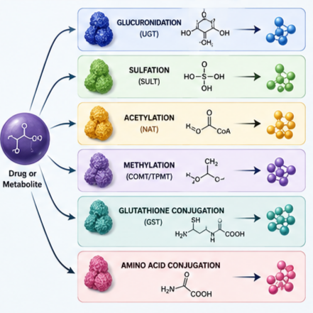
6.1 Glucuronidation
Glucuronidation is quantitatively the most significant Phase II reaction in humans, accounting for the elimination of approximately 30–50% of all drugs.
Mechanism:
UGT enzymes transfer glucuronic acid from UDP-glucuronic acid to the substrate. The reaction occurs at the luminal side of the endoplasmic reticulum.
Functional Groups:
– Alcohols (phenolic, primary, secondary, tertiary)
– Carboxylic acids (forming acyl glucuronides)
– Amines (forming N-glucuronides)
– Thiols (forming S-glucuronides)
Properties of Glucuronides:
– Highly hydrophilic, facilitating urinary or biliary excretion
– Generally biologically inactive (though some glucuronides retain activity or can be hydrolyzed back to active parent drug)
– Molecular weight > 300 Da promotes biliary excretion
Clinical Examples:
– Morphine: Forms morphine-3-glucuronide (inactive) and morphine-6-glucuronide (active analgesic)
– Acetaminophen: Glucuronidation is a major elimination pathway
– Bilirubin: Conjugation to bilirubin diglucuronide for biliary excretion
Enterohepatic Recirculation:
Glucuronides can be excreted in bile and deconjugated by intestinal β-glucuronidases, allowing reabsorption of the parent drug and prolonging its pharmacological effect.
6.2 Sulfation
Sulfation (also called sulfonation) is a high-affinity, low-capacity system that complements glucuronidation.
Mechanism:
SULT enzymes transfer a sulfate group from PAPS to hydroxyl or amino groups. The reaction occurs in the cytosol.
Properties of Sulfates:
– Highly hydrophilic, promoting urinary excretion
– Many sulfate conjugates are biologically inactive
– Sulfation is saturable at higher substrate concentrations
Clinical Examples:
– Acetaminophen: Sulfation accounts for 20–40% of clearance at therapeutic doses
– Steroids: Androgens, estrogens, and adrenal steroids are sulfated for excretion
– Catecholamines: Dopamine and norepinephrine are conjugated with sulfate
Limitations:
Sulfation capacity is limited by PAPS availability and the relatively small pool of sulfotransferase enzymes. This makes sulfation more susceptible to substrate saturation and drug interactions compared to glucuronidation.
6.3 Acetylation
N-acetylation is an important pathway for drugs containing primary amine groups, including aromatic amines, hydrazines, and sulfonamides.
Mechanism:
NAT enzymes transfer an acetyl group from acetyl-CoA to the amine substrate. The reaction occurs in the cytosol.
Properties of Acetylated Metabolites:
– Acetylation generally produces less water-soluble metabolites
– Acetylation is a major determinant of individual variability in drug response
– Genetic polymorphism in NAT2 results in slow and rapid acetylator phenotypes
Clinical Examples:
– Isoniazid: Acetylation is the major metabolic pathway; slow acetylators have higher risk of toxicity
– Hydralazine: Acetylation affects antihypertensive response
– Sulfonamides: Acetylation influences drug half-life and tissue penetration
Genetic Polymorphism:
NAT2 polymorphisms are among the best-characterized pharmacogenetic variants, with significant clinical implications:
– Slow acetylators: Increased risk of isoniazid-induced peripheral neuropathy and hepatotoxicity; increased risk of sulfonamide hypersensitivity
– Rapid acetylators: Reduced efficacy of isoniazid in tuberculosis treatment; increased risk of drug-induced lupus from hydralazine
6.4 Methylation
Methylation involves the transfer of a methyl group from S-adenosylmethionine (SAM) to various substrates, including catechols, thiols, and amines.
Mechanism:
Methyltransferases catalyze methyl transfer, with S-adenosylhomocysteine (SAH) as the byproduct.
Key Methyltransferase Enzymes:
1. Thiopurine S-Methyltransferase (TPMT)
– Metabolizes thiopurine drugs (azathioprine, 6-mercaptopurine, 6-thioguanine)
– Genetic polymorphism: 10% of individuals have intermediate activity; 0.3% are deficient
– Deficient patients require dose reduction to 5–10% of normal
– Pre-treatment TPMT genotype/phenotype testing is standard of care
2. Catechol O-Methyltransferase (COMT)
– Metabolizes catecholamines (dopamine, norepinephrine, epinephrine)
– Inhibitors (entacapone, tolcapone) used in Parkinson’s disease
– Val158Met polymorphism affects enzyme activity by 3- to 4-fold
3. Thiol Methyltransferase (TMT)
– Metabolizes sulfur-containing compounds
– Substrates include captopril, D-penicillamine
Clinical Examples:
– Azathioprine: TPMT deficiency leads to severe myelosuppression
– Parkinson’s disease: COMT inhibition prolongs levodopa half-life
6.5 Glutathione Conjugation
Glutathione (GSH) conjugation is a critical defense mechanism against reactive electrophiles, protecting cells from oxidative stress and toxicity.
Mechanism:
GSTs catalyze the addition of glutathione (a tripeptide: glutamyl-cysteinyl-glycine) to electrophilic substrates, generally through nucleophilic attack by the thiol group of cysteine.
Properties of Glutathione Conjugates:
– Highly hydrophilic, promoting excretion as mercapturic acids
– Often represents a detoxification pathway for reactive intermediates
– Can occasionally lead to bioactivation and toxicity
Clinical Significance:
– Acetaminophen toxicity: NAPQI (reactive metabolite) is detoxified by GSH conjugation. Overdose depletes GSH, leading to hepatotoxicity
– Chemotherapy resistance: Overexpression of GSTs in tumors can confer resistance to alkylating agents
– Environmental toxins: GSH protects against carcinogens, pesticides, and heavy metals
Glutathione Depletion:
Conditions that deplete GSH (e.g., acetaminophen overdose, chronic liver disease, poor nutrition) increase susceptibility to drug-induced toxicity.
6.6 Amino Acid Conjugation
Amino acid conjugation involves the formation of amide bonds between carboxylic acid drugs and endogenous amino acids, primarily glycine and glutamine.
Mechanism:
The process involves two steps:
1. Activation of the carboxylic acid to an acyl-CoA derivative (acyl-CoA synthetase)
2. Transfer of the acyl group to the amino acid (amino acid N-acyltransferase)
Clinical Examples:
– Salicylic acid: Glycine conjugation produces salicyluric acid (major elimination pathway)
– Nicotinic acid: Glycine conjugation to nicotinuric acid
– Ibuprofen: Amino acid conjugation is a minor pathway
Clinical Significance:
– Amino acid conjugation is generally a minor metabolic pathway for most drugs
– Less affected by genetic polymorphism compared to acetylation or methylation
– May be more clinically relevant in patients with impaired urea cycle or amino acid metabolism
7. Cofactors Required for Phase II Metabolism
Phase II conjugation reactions require high-energy cofactors that provide the conjugating moiety. These cofactors are essential for the activity of transferase enzymes and their availability can influence drug metabolism rates.
Table 2: Phase II Cofactors
| Enzyme Family | Cofactor | Conjugating Moiety | Source |
|---|---|---|---|
| UGTs | UDP-glucuronic acid | Glucuronic acid | Glucose metabolism |
| SULTs | PAPS (3′-phosphoadenosine-5′-phosphosulfate) | Sulfate | Sulfur amino acids |
| NATs | Acetyl-CoA | Acetyl | Carbohydrate metabolism |
| GSTs | Glutathione (GSH) | Glutathione | Amino acids (glutamate, cysteine, glycine) |
| MTs | S-Adenosylmethionine (SAM) | Methyl | Methionine |
صحت اور خوبصورتی کے مفید مشورے | Health & Beauty Tips : Skin Care، Hair Care، Weight Management، Healthy Lifestyle، Nutrition، Fitness اور Beauty Tips کے لیے وزٹ کریں۔ روزمرہ زندگی کو صحت مند اور خوبصورت بنانے کے لیے مفید اور معلوماتی آرٹیکلز پڑھیں۔
8. Transporters in Phase II Metabolism
Phase II metabolites, due to their increased polarity, require transporter proteins to cross biological membranes for excretion. Drug transporters are increasingly recognized as critical determinants of metabolite disposition and drug–drug interactions.
8.1 Hepatic Transporters
Canalicular (Biliary) Transporters:
– P-glycoprotein (P-gp): Exports glucuronides, sulfate conjugates into bile
– BCRP (Breast Cancer Resistance Protein): Mediates biliary excretion of sulfates and glucuronides
– MRP2 (Multidrug Resistance-Associated Protein 2): Primary transporter for glucuronide conjugates
– BSEP (Bile Salt Export Pump): Not a direct metabolite transporter, but may be inhibited by metabolite accumulation, leading to cholestasis
Basolateral (Sinusoidal) Transporters:
– MRP3 and MRP4: Efflux anions (glucuronides, sulfates) from hepatocytes into sinusoidal blood
– OATP1B1/1B3 (Organic Anion Transporting Polypeptides): Re-uptake of glucuronides from blood back into hepatocytes (“hepatocyte hopping”)
8.2 Renal Transporters
Uptake into Proximal Tubule:
– OAT1/3 (Organic Anion Transporters): Mediate uptake of anionic metabolites (glucuronides, sulfates) from blood
– OCT2 (Organic Cation Transporter 2): Transport of cationic metabolites
Secretion into Urine:
– MRP2, MRP4: Efflux into tubular lumen
– MATE1/2-K (Multidrug and Toxin Extrusion): Secretion of cationic drugs
8.3 Intestinal Transporters
– P-gp, BCRP, MRP2: Efflux back into intestinal lumen
– MRP3: Efflux from enterocytes into blood
8.4 Clinical Relevance
Transporter-mediated disposition of Phase II metabolites has important clinical implications:
Enterohepatic recirculation refers to the biliary excretion of glucuronides, followed by intestinal deconjugation and reabsorption of the parent drug. This can significantly prolong drug half-life.
Drug–drug interactions:
– Transporters are increasingly recognized as sites for clinically important drug interactions
– Approximately 2% of CYP-mediated interactions cannot be predicted from parent drug alone, highlighting the role of metabolites
– Inhibition of MRP2 can lead to metabolite accumulation and toxicity
– Modulation of intestinal transporters can alter systemic exposure
Regulatory considerations:
– Regulatory guidance (FDA, EMA) recommends characterization of metabolites that exceed 25% of parent drug systemic exposure or >10% of total drug-related radioactivity in safety testing
– Major excretory metabolites may warrant transporter interaction assessment even without systemic exposure
9. Pharmacokinetics of Phase II Reactions
9.1 Clearance
Phase II metabolism significantly contributes to total drug clearance, often representing the rate-limiting step for drug elimination.
Hepatic Clearance:
– High extraction ratio: Glucuronidation; e.g., morphine
– Low extraction ratio: Sulfation, acetylation; e.g., isoniazid
Renal Clearance:
– Glomerular filtration: Phase II metabolites (polar, low protein binding)
– Active secretion: Proximal tubule OATs for anionic metabolites
– Urinary pH: May influence reabsorption of uncharged metabolites
9.2 Enterohepatic Recirculation
Enterohepatic recirculation is a major feature of glucuronide metabolism:
1. Drug is absorbed → metabolized in liver to glucuronide
2. Glucuronide excreted in bile
3. Intestinal β-glucuronidases deconjugate the metabolite
4. Parent drug reabsorbed from intestine
5. Drug re-enters systemic circulation
This cycle can significantly prolong the pharmacological effect of drugs such as:
– Morphine: Morphine-6-glucuronide contributes to analgesic effect
– Estrogens: Enterohepatic recirculation contributes to the contraceptive effect of oral contraceptives
– Mycophenolate mofetil: Mycophenolic acid glucuronide is extensively recirculated
9.3 Saturation Kinetics
Many Phase II enzymes exhibit saturable kinetics (Michaelis-Menten). Saturation can occur at therapeutic doses for specific drugs or substrates.
Sulfation (low capacity, high affinity):
– Saturable at lower substrate concentrations
– Particularly evident for acetaminophen and catecholamines
– Overload of sulfation capacity can lead to increased toxicity or substrate competition
Glucuronidation (high capacity, variable affinity):
– Higher capacity than sulfation
– Saturation less common at therapeutic doses
– Can be overwhelmed at high doses (e.g., acetaminophen overdose)
Clinical Implications:
– Saturation leads to nonlinear pharmacokinetics
– Dose-dependent changes in plasma concentrations
– Increased risk of toxicity at high doses
10. Genetic Polymorphisms in Phase II Enzymes
Genetic variation in Phase II drug-metabolizing enzymes significantly influences interindividual differences in drug response, toxicity risk, and disease susceptibility.
Table 3: Pharmacogenetic Variants in Phase II Enzymes
| Enzyme | Gene | Polymorphism | Clinical Consequence |
|---|---|---|---|
| UGT1A1 | UGT1A1 | UGT1A1*28 (TA repeat) | Gilbert’s syndrome; reduced irinotecan clearance → myelotoxicity |
| UGT2B17 | UGT2B17 | Gene deletion | Reduced androgen metabolism; risk for cancer |
| NAT2 | NAT2 | Multiple (NAT2*4, *5, *6, *7) | Slow/rapid acetylator phenotype → isoniazid toxicity or failure |
| TPMT | TPMT | Multiple (TPMT*2, *3A, *3C) | Reduced thiopurine metabolism → severe myelosuppression |
| COMT | COMT | Val158Met | 3-4 fold enzyme activity variation; Parkinson’s disease risk |
| GSTP1 | GSTP1 | I105V | Reduced detoxification; cancer risk; chemotherapy resistance |
10.1 UGT1A1 Polymorphism and Irinotecan Toxicity
The UGT1A1 gene contains a polymorphic TA repeat in the TATA box (UGT1A1*28). Individuals homozygous for this variant have significantly reduced UGT1A1 activity.
Clinical Impact:
– Irinotecan is activated to SN-38 (active metabolite)
– SN-38 is detoxified by UGT1A1 glucuronidation
– Reduced UGT1A1 activity → SN-38 accumulation → increased toxicity (diarrhea, myelosuppression)
– Guidelines recommend UGT1A1 genotyping before irinotecan therapy
10.2 NAT2 Acetylation Polymorphism
The NAT2 gene exhibits extensive polymorphism with over 30 identified variants. The most common alleles determine the “slow” or “rapid” acetylator phenotype.
Clinical Impact:
– Slow acetylators: Increased risk of isoniazid-induced hepatotoxicity and peripheral neuropathy; increased risk of hydralazine-induced lupus
– Rapid acetylators: Reduced efficacy of isoniazid; increased risk of bladder cancer from arylamine exposure
10.3 TPMT Deficiency and Thiopurine Toxicity
TPMT deficiency is a well-characterized pharmacogenetic trait with significant clinical implications.
Clinical Impact:
– 0.3% of individuals are TPMT deficient; 10% have intermediate activity
– Azathioprine, 6-mercaptopurine, and 6-thioguanine are substrates
– Deficient patients develop severe myelosuppression at standard doses
– Dose reduction to 5–10% of standard recommended for deficient patients
– Pre-treatment TPMT genotyping or phenotyping is standard of care
11. Factors Affecting Phase II Metabolism
11.1 Age-Related Changes
Neonates and Infants:
– UGT activity is reduced in the neonatal period (particularly UGT1A1)
– Glucuronidation capacity reaches adult levels by 3–6 months
– Infants have increased risk of drug toxicity from drugs requiring glucuronidation (e.g., chloramphenicol)
Elderly:
– Generally, Phase II enzyme activity is less affected than Phase I
– Reduced liver mass and hepatic blood flow
– Decreased renal clearance of conjugated metabolites
– Increased risk of adverse effects from drugs with high hepatic clearance
11.2 Pregnancy
Pregnancy significantly alters drug metabolism:
– UGT isoforms are induced or repressed during pregnancy
– UGT1A4 (primary glucuronidator of lamotrigine) activity increases → lamotrigine dose increase may be required
– Sulfation and methylation may also be altered
– Changes are hormone-mediated (estrogens, progesterone)
11.3 Liver Disease
Cirrhosis:
– Impaired hepatic function reduces clearance of UGT substrates
– Decreased glucuronidation capacity for many drugs
– Impaired biliary excretion of conjugates
– Increased bioavailability of drugs with high first-pass metabolism
Hepatocellular Disease:
– Reduced enzyme expression in hepatocytes
– Impaired conjugation of bilirubin (UGT1A1)
– Increased risk of drug-induced hepatotoxicity
Cholestatic Disease:
– Impaired biliary excretion of glucuronides and other conjugates
– Accumulation of metabolites in systemic circulation
– May lead to metabolite toxicity or altered pharmacology
11.4 Kidney Disease
Renal Impairment:
– Glomerular filtration of polar metabolites is reduced
– Clearance of conjugated metabolites can be significantly impaired
– Risk of metabolite accumulation and toxicity
– Drugs requiring dose adjustment: Morphine (morphine-6-glucuronide accumulation → respiratory depression)
11.5 Drug–Drug Interactions
Phase II enzymes are subject to both induction and inhibition, although these mechanisms are less common than with Phase I enzymes.
Enzyme Induction:
– UGT induction: Rifampicin, phenobarbital, phenytoin → increased clearance of UGT substrates
– GST induction: Antioxidants, cruciferous vegetables
Enzyme Inhibition:
– UGT inhibition: Probenecid (inhibits UGT), valproic acid (inhibits UGT2B7)
– SULT inhibition: Non-steroidal anti-inflammatory drugs (NSAIDs) may inhibit SULT activity
Substrate Competition:
– Concurrent use of multiple drugs metabolized by the same enzyme
– Sulfation is particularly susceptible to competition due to limited PAPS availability
– High-dose acetaminophen can saturate sulfation pathway, leading to increased oxidative metabolism
11.6 Nutritional and Environmental Factors
Diet:
– Cruciferous vegetables (e.g., broccoli) induce GSTs
– Grapefruit juice may affect UGT enzymes (minor compared to CYP3A4)
– Protein-calorie malnutrition can reduce enzyme synthesis
Alcohol:
– Chronic alcohol consumption may affect UGT and GST activity
– Alcoholics are at increased risk of acetaminophen hepatotoxicity due to GSH depletion
Environmental Pollutants:
– Cigarette smoke induces GSTs and UGTs
– Pesticides and industrial chemicals can both induce and inhibit GSTs
12. Clinical Importance and Drug Examples
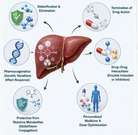
12.1 Drugs Primarily Undergoing Phase II Metabolism
Acetaminophen (Paracetamol):
– Major pathways: Glucuronidation (~50%), sulfation (~25–35%), oxidation (CYP450; ~5–15%)
– At therapeutic doses: Glucuronidation and sulfation are major pathways
– At toxic doses: Glucuronidation and sulfation become saturated → increased CYP450 oxidation → NAPQI formation → GSH depletion → hepatotoxicity
– Clinical pearl: N-acetylcysteine (NAC) replenishes GSH to prevent/limit hepatotoxicity
Morphine:
– Major pathways: Glucuronidation (UGT2B7) to morphine-3-glucuronide (inactive) and morphine-6-glucuronide (active)
– Renal impairment: Morphine-6-glucuronide accumulates → increased opioid effect (respiratory depression)
– Clinical pearl: Dose adjustment required in renal failure
Isoniazid:
– Major pathway: Acetylation (NAT2)
– Slow acetylators: Increased risk of toxicity (hepatotoxicity, peripheral neuropathy)
– Rapid acetylators: Reduced efficacy
– Clinical pearl: Pyridoxine supplementation prevents neuropathy, especially in slow acetylators
Azathioprine and 6-Mercaptopurine:
– Major pathway: Methylation (TPMT)
– TPMT deficiency: Severe myelosuppression at standard doses
– Clinical pearl: TPMT genotyping/phenotyping recommended before initiating therapy
Bilirubin (endogenous substrate):
– Conjugation: UGT1A1 glucuronidation
– Deficiency: Gilbert’s syndrome (mild unconjugated hyperbilirubinemia)
– Severe deficiency: Crigler-Najjar syndrome type I/II
12.2 Clinical Case Examples
Case 1: Isoniazid and NAT2 Polymorphism
A 45-year-old woman with active tuberculosis is started on isoniazid, rifampicin, ethambutol, and pyrazinamide. Two months later, she develops nausea, vomiting, and jaundice. Liver function tests show elevated transaminases and bilirubin.
Analysis:
– Isoniazid is primarily metabolized by NAT2
– Slow acetylator phenotype leads to isoniazid accumulation and hepatotoxicity
– Approximately 50% of the population has the slow acetylator phenotype (variable by ethnicity)
– Management: Stop isoniazid; consider alternative regimen (e.g., levofloxacin or other agents); pyridoxine supplementation to prevent neuropathy in slow acetylators
Case 2: Azathioprine and TPMT Deficiency
A 28-year-old man with Crohn’s disease is started on azathioprine 2 mg/kg/day. After two weeks, he develops severe pancytopenia. Bone marrow biopsy reveals severe marrow suppression.
Analysis:
– Azathioprine is metabolized by TPMT
– The patient likely has TPMT deficiency
– TPMT deficiency prevalence: ~0.3% (homozygous)
– Management: Stop azathioprine; consider alternative immunosuppressive therapy; TPMT genotyping should have been performed prior to initiation
Case 3: Irinotecan and UGT1A1 Polymorphism
A 72-year-old man with metastatic colon cancer is treated with irinotecan-based chemotherapy. After the first cycle, he develops severe diarrhea and febrile neutropenia.
Analysis:
– Irinotecan is activated to SN-38
– SN-38 is glucuronidated by UGT1A1
– Patients homozygous for UGT1A1*28 have reduced clearance → SN-38 accumulation → toxicity
– Management: Reduced irinotecan dose for subsequent cycles; consider UGT1A1 genotyping
13. Toxicology and Drug-Induced Liver Injury
13.1 Role of Phase II Metabolism in Toxicity
While Phase II reactions are generally detoxifying, conjugation can lead to toxicity through several mechanisms:
1. Bioactivation via Glutathione Conjugation:
– Glutathione conjugates can be further metabolized to reactive species
– Classic example: Nephrotoxicity of hydroquinone and bromobenzene involves quinone-glutathione conjugates leading to cellular damage
– 1,2-Dibromo-3-chloropropane bioactivation via GSTs causes kidney and testicular toxicity
2. Glucuronide-Mediated Toxicity:
– Acyl glucuronides are chemically reactive and can bind to proteins
– Some acyl glucuronides are toxic (e.g., diclofenac, valproic acid)
– The “metabolic shift” of some drugs from glucuronidation to oxidative metabolism in toxicity
3. Formation of Reactive Intermediates:
– Certain Phase II metabolites are reactive and form covalent bonds with cellular macromolecules
– Example: N-acetylation of aromatic amines to form reactive intermediates
– Example: Sulfation of some drugs to form sulfate esters that can react with DNA (bioactivation)
4. Acetaminophen Hepatotoxicity:
– The classic example of Phase II metabolism failure leading to toxicity:
– Therapeutic doses: Glucuronidation + sulfation clear acetaminophen
– Overdose: Glucuronidation + sulfation saturated → increased CYP450 oxidation → NAPQI (reactive metabolite)
– GSH depletion → NAPQI binds to cellular proteins → hepatocellular damage
13.2 Drug-Induced Liver Injury (DILI)
Drug-induced liver injury is a major clinical concern in drug development and clinical practice. Phase II metabolism can influence DILI risk:
Contributing Factors:
– Genetic polymorphisms in Phase II enzymes (NAT2, UGTs, GSTs) increasing susceptibility
– Cofactor depletion (GSH depletion in acetaminophen toxicity)
– Formation of reactive conjugates (acyl glucuronides)
– Drug–drug interactions altering Phase II metabolism
Examples of DILI Associated with Phase II Metabolism:
– Isoniazid: Hepatotoxicity associated with NAT2 slow acetylator phenotype
– Valproic acid: Acyl glucuronide formation linked to hepatotoxicity
– Acetaminophen: GSH depletion and reactive metabolite formation
– Ticlopidine: Sulfation to reactive metabolite linked to hepatotoxicity
Common Misconceptions
Misconception 1: Phase I Metabolism Must Precede Phase II Metabolism
Reality: While sequential metabolism (Phase I then Phase II) is common, many drugs undergo direct Phase II conjugation if they contain suitable functional groups (e.g., morphine, acetaminophen).
Misconception 2: Phase II Reactions Always Produce Inactive Metabolites
Reality: While most Phase II metabolites are inactive, notable exceptions exist. Morphine-6-glucuronide is pharmacologically active (more potent than morphine itself). Some glucuronides are cleaved to the active parent drug via enterohepatic recirculation.
Misconception 3: Phase II Metabolism is Always Detoxifying
Reality: Phase II reactions are generally detoxifying, but exceptions include:
– Bioactivation of prodrugs
– Formation of toxic conjugates (e.g., acyl glucuronides)
– Glutathione conjugation leading to reactive intermediates
Misconception 4: Phase II Metabolism is Less Important Than Phase I
Reality: Phase II metabolism accounts for the elimination of approximately 30–50% of all drugs. In many cases (e.g., acetaminophen, morphine, azathioprine), Phase II reactions represent the major or sole elimination pathway.
Misconception 5: Genetic Polymorphisms in Phase II Enzymes Are Rare
Reality: Genetic polymorphisms are relatively common in Phase II enzymes:
– NAT2 slow acetylator phenotype: 50% of population (variable by ethnicity)
– TPMT deficiency: 10% with intermediate activity
– UGT1A1*28 homozygous: 10% of Caucasians
Question . What is Phase II drug metabolism?
Question . What are the major Phase II reactions?
Question . Why is Phase II metabolism important?
Question . What enzymes catalyze Phase II reactions?
Question . Where does Phase II metabolism occur?
Question . What is the difference between Phase I and Phase II metabolism?
Question . What is enterohepatic recirculation?
Question . What is the clinical significance of NAT2 polymorphism?
Question . How does age affect Phase II metabolism?
Question . How do drug–drug interactions affect Phase II metabolism?
Question . Can Phase II metabolism activate drugs?
Question . Why is TPMT testing recommended before azathioprine?
Question . What is the role of glutathione in acetaminophen toxicity?
Question . What is the clinical impact of UGT1A1 polymorphism?
Question . How does renal impairment affect Phase II metabolism?
Question . What are the advantages of glucuronidation over sulfation?
Question . What is the role of transporters in Phase II metabolism?
Question . Which drugs are primarily metabolized by Phase II reactions?
Question . What is the clinical significance of methylation in drug metabolism?
Question . How does liver disease affect Phase II metabolism?
Question . What is the role of GSTs in detoxification?
Question . How does pregnancy affect Phase II metabolism?
Question . What is amino acid conjugation?
Question . Why is glucuronidation considered the most important Phase II reaction?
Question . How is Phase II metabolism regulated?
Question . What is the role of COMT in Parkinson’s disease?
Question . How does malnutrition affect Phase II metabolism?
Question . What is the clinical significance of acetylation polymorphism?
Question . Can Phase II metabolites be pharmacologically active?
Question . What is the fate of Phase II conjugates?
Question . How does smoking affect Phase II metabolism?
Question . What is the relationship between Phase II metabolism and personalized medicine?
Question . How does sulfation differ from glucuronidation?
Question . What is the role of Phase II metabolism in drug development?
Question . What are acyl glucuronides and why are they clinically relevant?
Question . What are the genetic polymorphisms of TPMT?
Question . How does enterohepatic recirculation affect drug half-life?
Question . What is the role of Phase II metabolism in cancer chemotherapy?
Question . Can Phase II metabolism be induced?
Question . How does obesity affect Phase II metabolism?
Question . What is the role of MRP transporters in Phase II metabolism?
Question . Why is bilirubin conjugation clinically important?
Question . Can Phase II metabolism lead to drug resistance?
Question . How does the intestine contribute to Phase II metabolism?
Question . What is the clinical significance of methylation polymorphisms?
Question . How does Phase II metabolism affect drug clearance in renal failure?
Question . What is the role of OATP transporters in Phase II metabolism?
Question . Are there any drugs that undergo both Phase I and Phase II metabolism?
Question . How does Phase II metabolism affect drug–food interactions?
Question . What is the future of Phase II metabolism research?
Key Takeaways
1. Phase II metabolism (conjugation) is a critical detoxification pathway that converts lipophilic drugs into hydrophilic, excretable conjugates through the action of transferase enzymes.
2. Major conjugation reactions include glucuronidation, sulfation, acetylation, methylation, glutathione conjugation, and amino acid conjugation. Glucuronidation is quantitatively the most significant.
3. Key enzymes involved are UGTs, SULTs, NATs, GSTs, and methyltransferases, each with distinct substrate specificity, tissue distribution, and regulatory mechanisms.
4. Genetic polymorphisms in Phase II enzymes (e.g., UGT1A1, NAT2, TPMT) significantly influence drug efficacy, toxicity risk, and therapeutic outcomes, making pharmacogenetic testing increasingly important in clinical practice.
5. Drug transporters play a critical role in the disposition of Phase II metabolites, mediating their excretion in bile, urine, and intestine, and contributing to enterohepatic recirculation.
6. Phase II metabolism is not always detoxifying: Bioactivation via glutathione conjugation, acyl glucuronide formation, and production of reactive metabolites can lead to toxicity.
7. Clinical factors including age, pregnancy, liver and kidney disease, drug interactions, and nutritional status significantly affect Phase II metabolism, requiring careful dose adjustment and patient monitoring.
8. Regulatory guidelines (FDA, EMA) recommend characterization of major metabolites and their transport properties during drug development to assess drug–drug interaction potential and safety.
9. Personalized medicine applications of Phase II pharmacogenetics (e.g., TPMT, UGT1A1, NAT2 testing) are increasingly integrated into clinical practice to optimize drug selection and dosing.
10. Future research in Phase II metabolism focuses on better understanding genetic variants, transporter interactions, enterohepatic recirculation, and developing more precise models for predicting drug disposition in diverse patient populations.
References
Textbooks and Major References
1. Goodman & Gilman’s The Pharmacological Basis of Therapeutics, 13th Edition. Chapter: Drug Metabolism. (General reference for drug metabolism principles)
2. Katzung BG, Vanderah TW. Basic & Clinical Pharmacology, 15th Edition. Chapter: Pharmacokinetics & Pharmacodynamics. (General reference for pharmacokinetic principles)
3. Rang HP, Dale MM, Ritter JM, Flower RJ. Rang and Dale’s Pharmacology, 9th Edition. Chapter: Pharmacokinetics. (General reference for drug metabolism)
4. Murray M. Chapter 6B. Phase II Drug-Metabolizing Enzymes. In: AccessPharmacy, McGraw-Hill Medical.
5. Ashton M, Groundwater PW, Stocker S, Todd A. An Integrated Guide to Human Drug Metabolism: From Basic Chemical Transformations to Drug-Drug Interactions. Elsevier, 2024.
Peer-Reviewed Journal Articles
6. Jancova P, et al. Phase II drug metabolizing enzymes. Biomed Pap Med Fac Univ Palacky Olomouc Czech Repub. 2010;154(2):103-116.
7. Testa B, Krämer SD. The biochemistry of drug metabolism–an introduction: part 4. reactions of conjugation and their enzymes. Chem Biodivers. 2008;5(11):2171-2336.
8. Zamek-Gliszczynski MJ, et al. Understanding the transport properties of metabolites: Case studies and considerations for drug development. Drug Metab Dispos. 2014;42(4):650-664.
9. Murray M. Pharmacogenetics of phase I and phase II drug metabolism. Curr Pharm Des. 2010;16(2):204-215.
10. Drug Metabolism: Phase II Enzymes. In: Comprehensive Pharmacology. Elsevier, 2022:563-584.
11. Phase II enzymes and bioactivation. Colloquium summary. 1995.
12. Drug Metabolism: Phase II Reactions. JoVE Science Education Database.
13. Drug Metabolism Synthetic (Phase II) Reactions. In: The ADME Encyclopedia. Springer Nature, 2021.
14. Metabolism of Drugs and Xenobiotics. In: Reference Module in Biomedical Sciences. Elsevier.
15. Drug Metabolism: Phase II Reactions. CORE.
Regulatory and Clinical Guidelines
16. FDA Guidance for Industry: Drug Interaction Studies – Study Design, Data Analysis, Implications for Dosing, and Labeling Recommendations. (2012, 2020).
17. EMA Guideline on the Investigation of Drug Interactions. (2012, 2021).
18. FDA Guidance for Industry: Safety Testing of Drug Metabolites. (2008, updated 2020).
19. ICH Guideline M3(R2) on Nonclinical Safety Studies for the Conduct of Human Clinical Trials and Marketing Authorization for Pharmaceuticals. (2009).
20. Clinical Pharmacogenetics Implementation Consortium (CPIC) Guidelines: TPMT, UGT1A1, NAT2, COMT genotyping recommendations.
21. Lexicomp Drug Interactions. Various monographs, 2024. (General reference for drug–drug interactions)
22. AHFS Clinical Drug Information. Various monographs, 2024. (General reference for drug metabolism information)
23. NCBI Bookshelf: StatPearls – Drug Metabolism. (General reference for drug metabolism)
24. WHO Model List of Essential Medicines. 2023. (General reference)
25. NIH National Library of Medicine: LiverTox – Clinical and Research Information on Drug-Induced Liver Injury. (General reference for toxicity)
Last Updated: This article was prepared based on the latest available evidence and guidelines. Please check for updates in clinical practice recommendations.
Medical Disclaimer ; This article is for educational and informational purposes only and does not constitute medical advice. The content is intended for healthcare professionals, medical students, and researchers. Clinical decisions must be made by qualified healthcare providers based on individual patient circumstances. Drug dosing, interactions, and therapeutic strategies should follow official prescribing information and clinical guidelines. The author and publisher disclaim any liability for any adverse effects arising from the use or application of information contained herein.