Zero-Order Kinetics Made Easy 12 Must-Know Clinical Concepts with Real Drug Examples
Clinical Scenario
Consider a 45-year-old man brought to the emergency department with altered mental status, slurred speech, and ataxic gait. His friends report he has been drinking heavily for the past six hours at a wedding celebration. The emergency resident calculates that his blood ethanol concentration is approximately 220 mg/dL and estimates, based on zero-order elimination kinetics, that it will take roughly 11 to 14 hours for his blood alcohol to reach legally acceptable levels for safe discharge. The resident understands something fundamental about ethanol metabolism: unlike most drugs that are eliminated as a constant percentage per unit time, ethanol disappears from the body at a constant rate regardless of its concentration. This is the essence of zero-order kinetics, a concept that carries profound implications for drug dosing, toxicity prediction, and therapeutic drug monitoring across numerous medications encountered in clinical practice.
This scenario illustrates why understanding zero-order kinetics is not merely an academic exercise in memorizing pharmacokinetic equations. It directly informs clinical decision-making, influences patient safety, and explains why certain drugs become dangerous when their metabolic pathways are overwhelmed. Throughout this comprehensive review, we will explore the biochemical principles, mathematical foundations, clinical applications, and therapeutic implications of zero-order elimination kinetics in a manner that bridges foundational pharmacology with bedside medicine.
What Is Zero-Order Kinetics?
 A Foundational Definition
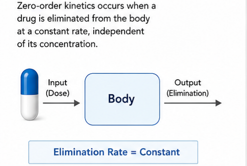
Zero-order kinetics, in the context of clinical pharmacology, describes a pattern of drug elimination where the rate of removal from the body remains constant and independent of the drug’s plasma concentration. In practical terms, this means that a fixed amount of drug is metabolized or eliminated per unit time, rather than a fixed percentage. When a drug follows zero-order kinetics, doubling the dose does not double the elimination rate; instead, the same absolute quantity is processed regardless of how much drug is present in the body.
This phenomenon is also referred to as saturation kinetics because it occurs when the enzymatic systems responsible for drug metabolism or the transport mechanisms involved in elimination become fully saturated. Once saturation occurs, the metabolic machinery operates at its maximum capacity, or Vmax, and cannot increase its rate of drug processing even when presented with higher substrate concentrations. The clinical consequence is profound: drugs that exhibit zero-order kinetics have no true half-life in the traditional sense, and small increases in dosage can lead to disproportionately large increases in plasma concentration, dramatically elevating the risk of toxicity.
Understanding zero-order kinetics requires recognizing that it represents a departure from the more commonly encountered first-order elimination pattern. In first-order kinetics, which governs the elimination of most therapeutic agents, a constant fraction or percentage of the drug is removed per unit time, making the elimination rate directly proportional to the drug concentration. This proportionality provides a safety buffer, as higher concentrations lead to faster elimination. Zero-order kinetics removes this buffer entirely, creating a pharmacokinetic environment where accumulation can occur rapidly and unpredictably.
Historical Development of Zero-Order Kinetics in Pharmacology
The recognition of saturation kinetics in drug metabolism emerged gradually through clinical observations and experimental studies conducted throughout the twentieth century. The mathematical foundation for understanding enzyme saturation was established by Leonor Michaelis and Maud Menten in 1913, who described the kinetic behavior of enzymes acting on substrates. Their work demonstrated that enzymatic reactions proceed at rates proportional to substrate concentration only at low concentrations, eventually reaching a maximum velocity when all enzyme active sites are occupied. This Michaelis-Menten relationship would later prove essential for understanding why certain drugs exhibit dose-dependent kinetics.
The clinical significance of zero-order elimination became apparent in the 1930s and 1940s when researchers studying ethanol metabolism observed that alcohol disappeared from the blood at a constant rate regardless of its concentration. Erik Widmark, a Swedish physician and researcher, conducted pioneering studies on ethanol pharmacokinetics in the 1920s and 1930s, developing mathematical models that described the linear decline of blood alcohol concentrations over time. His work established that the average person eliminates approximately 10 to 15 milligrams of ethanol per 100 milliliters of blood per hour, a value that has informed clinical and forensic practice for nearly a century.
The recognition that phenytoin exhibits saturable metabolism within the therapeutic range emerged during the 1960s, when clinicians observed that small dosage adjustments in epileptic patients sometimes produced dramatic changes in serum concentrations and clinical effects. This observation led to the characterization of phenytoin as a drug with Michaelis-Menten elimination kinetics, fundamentally altering approaches to dosing and therapeutic drug monitoring for this anticonvulsant. Subsequent research identified salicylates, theophylline in certain circumstances, and several other agents as exhibiting dose-dependent elimination, expanding the clinical relevance of zero-order kinetic principles.
The development of therapeutic drug monitoring as a clinical discipline owes much to the recognition of saturation kinetics, as it became clear that drugs with nonlinear elimination characteristics required careful concentration monitoring to avoid toxicity while maintaining efficacy. Modern pharmacokinetic modeling incorporates these principles, allowing clinicians to predict concentration changes and individualize dosing regimens based on patient-specific factors and measured drug levels.
Basic Principles of Drug Elimination
 First-Order and Zero-Order Processes
Drug elimination encompasses all processes that remove an administered drug from the body, including metabolism in the liver and other tissues, renal excretion of unchanged drug and metabolites, biliary secretion, and pulmonary elimination for volatile agents. The kinetic behavior of these elimination processes determines how drug concentrations change over time and directly influences dosing regimens, safety margins, and therapeutic outcomes.
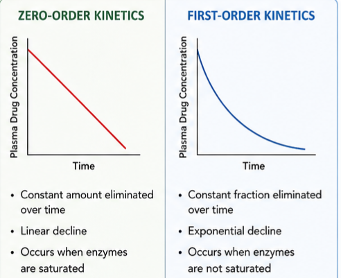
In first-order elimination kinetics, the rate of drug removal is directly proportional to the drug concentration in plasma. When the plasma concentration is high, the absolute amount of drug eliminated per unit time is large; as the concentration falls, the elimination rate decreases proportionally. This relationship produces the characteristic exponential decay curve observed with most medications, where a constant fraction of the remaining drug is eliminated during each successive time interval. The practical expression of this principle is the drug half-life, which represents the time required for the plasma concentration to decrease by fifty percent. Critically, the half-life remains constant for drugs following first-order kinetics, regardless of the starting concentration, making it a reliable parameter for predicting drug accumulation and washout.
The mathematical expression for first-order elimination states that the rate of change in drug amount equals the elimination rate constant multiplied by the amount of drug present. Integration of this differential equation yields the exponential decay function that describes concentration-time profiles for first-order drugs. The elimination rate constant, typically denoted as k or kel, has units of reciprocal time and is related to the half-life by a simple logarithmic relationship.
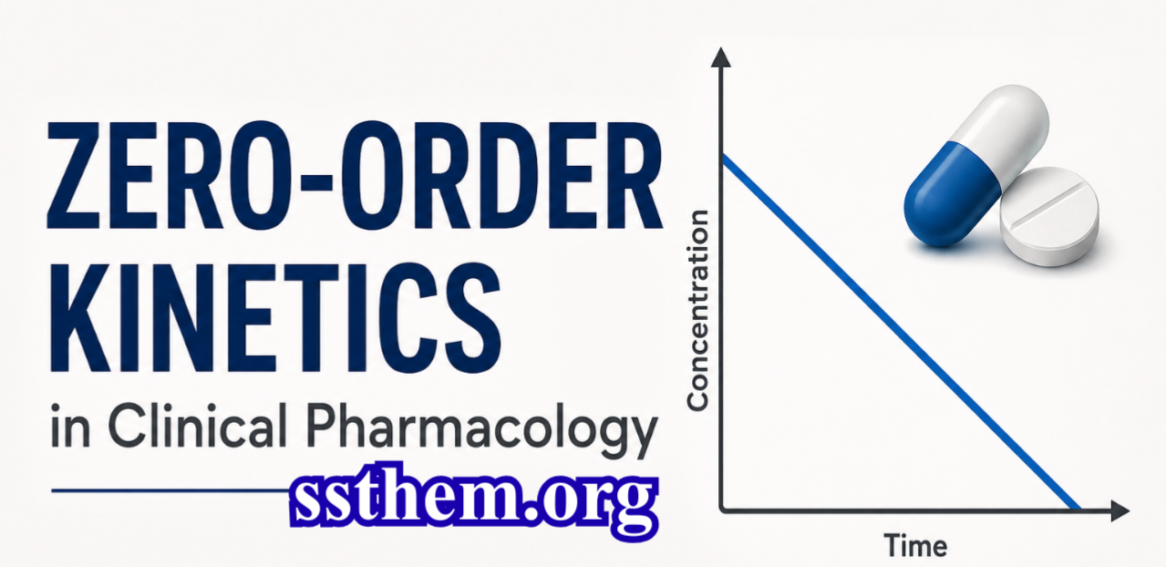
Zero-order elimination kinetics operate under fundamentally different principles. When elimination follows zero-order behavior, the rate of drug removal is constant and independent of plasma concentration. The metabolic or excretory capacity operates at its maximum, and the system removes a fixed quantity of drug per unit time regardless of how much drug remains. This produces a linear decline in drug concentration over time when plotted on standard arithmetic coordinates, rather than the exponential decline characteristic of first-order processes.
It is essential to recognize that zero-order kinetics typically arises from saturation of first-order processes. At low concentrations, the same drug may exhibit first-order elimination because the metabolic enzymes or transporters are not saturated. As concentrations increase beyond the capacity of these systems, the elimination pattern transitions from first-order to mixed-order and eventually to zero-order behavior. This concentration dependence explains why the same drug can display different elimination kinetics depending on the dose administered and the individual patient’s metabolic capacity.
The Mathematical Basis of Zero-Order Kinetics
The mathematical description of zero-order kinetics provides the quantitative framework necessary for predicting drug concentrations, designing dosing regimens, and understanding the clinical implications of saturation metabolism. The fundamental differential equation for zero-order elimination states that the rate of change in drug amount with respect to time equals a constant, typically expressed as negative Vmax, representing the maximum rate of elimination.
When integrated, this differential equation yields a linear function describing how drug concentration decreases over time. If C represents the drug concentration at any time t, and C0 represents the initial concentration, the concentration at any subsequent time equals the initial concentration minus the product of the zero-order elimination rate constant and the elapsed time. The zero-order rate constant carries units of mass per volume per time, reflecting that a fixed amount of drug is removed from each unit volume of distribution per unit time.
This linear relationship has several critical implications for clinical pharmacology. First, the time required to eliminate a given fraction of the drug depends on the initial concentration. Unlike first-order kinetics where the half-life remains constant, zero-order elimination provides no fixed half-life. The time needed to reduce the concentration from 100 mg per liter to 50 mg per liter is the same as the time needed to reduce it from 50 mg per liter to 0 mg per liter, assuming no further drug absorption occurs. Second, the elimination rate constant provides complete information about how quickly the drug will be removed, but this rate cannot be increased by increasing the drug concentration.
The clinical application of these mathematical principles becomes particularly important when calculating the duration of pharmacological effects or toxicity. For ethanol, with a typical elimination rate of 15 mg per deciliter per hour, a patient presenting with a blood alcohol concentration of 300 mg per deciliter will require approximately 20 hours to completely eliminate the alcohol from their system, assuming a zero-order process throughout. If the same patient had a concentration of 150 mg per deciliter, elimination would require approximately 10 hours. The linear relationship makes such calculations straightforward but also highlights the danger: if the initial concentration is high, elimination takes a predictably long time, and no physiological mechanism can accelerate this process.
Characteristics of Zero-Order Elimination: Identifying Features
Zero-order drug elimination exhibits several distinctive characteristics that allow clinicians to recognize when saturation kinetics are occurring and to anticipate the associated clinical implications. Understanding these features facilitates appropriate drug selection, dosing, and monitoring in clinical practice.
The most fundamental characteristic of zero-order elimination is the constant rate of drug removal irrespective of plasma concentration. When drug concentrations are measured serially and plotted against time on standard arithmetic coordinates, zero-order elimination produces a straight line with a constant negative slope. This linear decline contrasts sharply with the curved exponential decline observed with first-order kinetics. Clinicians familiar with interpreting drug concentration data can recognize zero-order behavior by this linear pattern, although confirming saturation kinetics typically requires measurements at multiple concentrations.
A second defining characteristic is the absence of a constant half-life. For drugs following first-order kinetics, the half-life is a fundamental and unchanging parameter that allows prediction of accumulation during multiple dosing and estimation of time to steady state. Zero-order drugs lack this convenient property entirely. The time required to eliminate fifty percent of the drug from the body depends on the starting concentration, with higher concentrations requiring proportionally longer elimination times. This variable half-life complicates dosing decisions and necessitates concentration monitoring rather than reliance on population pharmacokinetic parameters.
The third characteristic involves the relationship between dose and steady-state concentration. For first-order drugs, steady-state concentration increases proportionally with dose, meaning that doubling the maintenance dose ultimately doubles the average plasma concentration. For zero-order drugs, the relationship becomes nonlinear and unpredictable. At concentrations approaching or exceeding the metabolic capacity, small dose increases can produce disproportionately large increases in plasma concentration because the elimination system cannot increase its rate to match the increased input. This nonlinear dose-concentration relationship is the pharmacokinetic basis for the narrow therapeutic indices observed with several zero-order drugs.
A fourth characteristic is that zero-order elimination is typically concentration-dependent for a given drug. At low concentrations, before saturation occurs, the same drug usually follows first-order kinetics. The transition from first-order to zero-order behavior occurs when drug concentrations exceed the Michaelis constant for the metabolizing enzyme system, representing the concentration at which the elimination rate reaches half of its maximum value. This transitional behavior means that some drugs may exhibit first-order kinetics at therapeutic doses but shift toward zero-order elimination when doses are increased or when accumulation occurs due to impaired metabolism.
The Mechanism of Saturation Kinetics
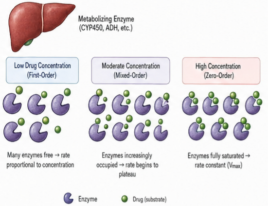 Enzyme Biology : The biochemical basis for zero-order drug elimination lies in the saturation of the enzymatic systems responsible for drug metabolism. Most drugs undergo biotransformation through enzyme-mediated reactions, predominantly catalyzed by the cytochrome P450 superfamily of heme-containing monooxygenases. These enzymes, like all biological catalysts, operate through the formation of enzyme-substrate complexes that proceed through transition states to yield products. The catalytic cycle involves substrate binding, electron transfer, oxygen activation, and product release, each step contributing to the overall kinetics of the reaction.
Enzyme molecules possess a finite number of active sites, and at any given moment, each enzyme molecule can process only one substrate molecule or a limited number of substrate molecules. When drug concentrations are low relative to the available enzyme, most active sites remain unoccupied at any instant, and the rate of product formation increases proportionally with substrate concentration. This represents the first-order region of the enzyme kinetic profile. As drug concentrations rise, an increasing fraction of enzyme active sites become occupied, and the rate of product formation approaches the maximum capacity of the enzyme population. Eventually, at sufficiently high substrate concentrations, essentially all enzyme active sites are occupied, and the reaction rate reaches its maximum, designated Vmax.
The Michaelis-Menten equation provides the mathematical framework describing this saturation behavior. The equation expresses reaction velocity as a function of substrate concentration, maximum velocity, and the Michaelis constant, which represents the substrate concentration at which the reaction velocity reaches half of the maximum. When the drug concentration is well below the Michaelis constant, the reaction rate approximates first-order behavior. When the drug concentration greatly exceeds the Michaelis constant, the reaction rate approaches zero-order behavior at Vmax. In the intermediate range, the kinetics are mixed, displaying characteristics of both first-order and zero-order processes.
The clinical translation of this enzymatic behavior is that drugs metabolized primarily through a single saturable pathway are candidates for zero-order kinetics when administered at doses that produce concentrations approaching or exceeding the Michaelis constant for that pathway. The specific cytochrome P450 isoforms involved, their expression levels in individual patients, and the presence of competing substrates or inhibitors all influence the concentration at which saturation occurs and the maximum elimination rate achievable.
The Michaelis-Menten Relationship in Clinical Pharmacokinetics
The Michaelis-Menten model, originally developed to describe single-substrate enzyme kinetics, provides the theoretical foundation for understanding dose-dependent drug elimination in clinical settings. This model reconciles the apparent contradiction that a drug can follow first-order kinetics under some circumstances and zero-order kinetics under others by describing elimination as a saturable process that transitions between kinetic orders depending on the drug concentration relative to the metabolic capacity.
The Michaelis-Menten equation describes the rate of drug elimination as a function of drug concentration. At very low concentrations relative to the Michaelis constant, the denominator of the equation is dominated by the constant term, making the elimination rate approximately proportional to concentration, consistent with first-order kinetics. At concentrations far exceeding the Michaelis constant, the concentration term in the denominator dominates, and the elimination rate approaches the maximum velocity, representing zero-order behavior. Between these extremes, the kinetics follow a mixed pattern that requires the full Michaelis-Menten expression for accurate description.
The clinical importance of the Michaelis constant lies in its relationship to therapeutic drug concentrations. When the therapeutic range of a drug is near or above the Michaelis constant for its primary elimination pathway, the drug will exhibit dose-dependent kinetics at clinically used doses. Phenytoin exemplifies this situation, with therapeutic concentrations of 10 to 20 mg per liter approaching or exceeding the typical Michaelis constant of 4 to 8 mg per liter for the CYP2C9-mediated hydroxylation pathway. Consequently, phenytoin displays nonlinear kinetics throughout much of its therapeutic range, with the degree of nonlinearity increasing at higher concentrations.
The maximum velocity parameter carries equal clinical significance, as it determines the absolute ceiling on drug elimination capacity. Factors that reduce enzyme expression or activity, such as genetic polymorphisms, hepatic disease, or concomitant medications that inhibit the relevant metabolic pathway, can substantially decrease Vmax. A patient with reduced Vmax will experience saturation at lower drug concentrations and will eliminate the drug more slowly at any given concentration, increasing the risk of accumulation and toxicity.
Understanding the Michaelis-Menten relationship enables clinicians to anticipate when dose adjustments may produce unexpected changes in drug concentrations. Near the saturation point, the slope of the concentration-response curve for dose increases becomes steep, meaning that a modest increase in daily dose can produce a disproportionately large increase in steady-state concentration. This nonlinearity necessitates cautious dose titration and, where available, therapeutic drug monitoring to guide therapy.
Why Zero-Order Kinetics Occurs: The Physiology of Metabolic Saturation
The transition from first-order to zero-order elimination occurs when the capacity of drug-metabolizing systems becomes limiting relative to the amount of drug requiring processing. This saturation can occur at multiple levels within the pharmacokinetic pathway, including enzymatic metabolism, active transport, protein binding, and blood flow delivery. Understanding the physiological bottlenecks that produce saturation kinetics provides insight into why specific drugs exhibit zero-order behavior and how patient factors modify the tendency toward saturation.
Enzymatic saturation represents the most common and clinically significant mechanism producing zero-order kinetics. The cytochrome P450 system, while possessing substantial metabolic capacity across its numerous isoforms, expresses each specific enzyme in limited quantities. CYP2E1, responsible for ethanol oxidation, is present in finite amounts in hepatocytes, and when ethanol intake exceeds the oxidative capacity of available enzyme, blood concentrations rise and elimination proceeds at a constant maximum rate. Similarly, CYP2C9, which catalyzes the para-hydroxylation of phenytoin, can become saturated at therapeutic phenytoin concentrations because the enzyme’s capacity is limited relative to typical dosing requirements.
Active transport saturation provides another mechanism for zero-order kinetics, particularly for drugs that undergo extensive renal tubular secretion or biliary excretion. These transport processes rely on carrier proteins such as organic anion transporters and organic cation transporters that exhibit saturable binding kinetics. At high drug concentrations, these transporters operate at maximum capacity, and further increases in concentration do not increase the rate of secretion. Methotrexate, penicillin derivatives, and certain cephalosporins can saturate renal tubular secretory mechanisms, though the clinical significance varies among these agents.
Protein binding saturation, while less commonly the primary cause of zero-order elimination kinetics, can contribute to nonlinear pharmacokinetic behavior. Drugs that are highly protein-bound occupy a finite number of binding sites on albumin or alpha-1 acid glycoprotein. When binding sites approach saturation, the free fraction of drug increases, potentially altering both the volume of distribution and the rate of elimination. However, the clinical impact of protein binding saturation on overall elimination kinetics is typically modest compared to metabolic or transport saturation.
Blood flow limitation represents a physiological ceiling on drug delivery to eliminating organs. For drugs with very high hepatic extraction ratios, clearance approaches hepatic blood flow, and elimination becomes dependent on delivery rather than on intrinsic metabolic capacity. While this produces flow-limited rather than capacity-limited kinetics, the practical consequence is similar: elimination rate becomes independent of drug concentration above a certain threshold. Understanding this distinction between capacity-limited and flow-limited elimination is important for interpreting pharmacokinetic data and predicting the effects of physiological changes or drug interactions.
Graphical Representation of Zero-Order Kinetics
The visual representation of drug concentration changes over time provides one of the most intuitive methods for understanding and recognizing zero-order elimination kinetics. Pharmacokinetic graphs serve both educational and clinical purposes, allowing rapid identification of elimination patterns and facilitating communication of kinetic principles to learners and colleagues.
When drug concentration is plotted on the vertical axis against time on the horizontal axis using standard arithmetic scales, zero-order elimination produces a straight line descending with a constant slope. This linear decline reflects the fundamental property of zero-order kinetics: the same absolute amount of drug is eliminated during each time interval, resulting in equal decrements in concentration when plotted arithmetically. The slope of this line equals the negative of the zero-order elimination rate constant, and extrapolation to the time axis provides an estimate of the time required for complete elimination from the initial concentration.
The same zero-order data plotted on semi-logarithmic coordinates, where the concentration axis uses a logarithmic scale while the time axis remains arithmetic, produces a curve with a distinctive shape. Rather than the straight line characteristic of first-order elimination on semi-logarithmic plots, zero-order data appear convex, with the downward slope becoming progressively steeper as concentration declines. This increasing steepness reflects that a constant absolute decrement represents an increasing fraction of the remaining drug as the total amount decreases. Familiarity with the appearance of zero-order data on both arithmetic and semi-logarithmic plots enables clinicians to recognize saturation kinetics when reviewing patient data or published pharmacokinetic studies.
The comparison between first-order and zero-order elimination on the same set of axes provides particular insight into their differences. On an arithmetic plot, the first-order curve shows an exponential decline that is initially steep but gradually flattens, never reaching zero concentration in finite time in theory. The zero-order line declines at a constant rate, reaching the axis at a predictable time. On a semi-logarithmic plot, the first-order data form a straight line whose slope relates to the elimination rate constant, while the zero-order data curve downward. These graphical distinctions are not merely academic; they provide the basis for determining which kinetic model applies to a particular drug in a particular patient and for calculating the pharmacokinetic parameters necessary for therapeutic decision-making.
The clinical utility of pharmacokinetic graphing extends to the visualization of dose-concentration relationships. When steady-state concentration is plotted against daily dose for a drug following first-order kinetics, the relationship is linear, with the slope determined by the drug’s clearance. For a drug exhibiting saturable metabolism, the dose-concentration curve is concave upward, demonstrating that concentration increases more than proportionally with dose. This graph explains visually why dose increases in the nonlinear range must be made cautiously and in small increments, as the steep portion of the curve predicts large concentration changes from small dosage adjustments.
Zero-Order Versus First-Order Kinetics: A Systematic Comparison
The distinction between zero-order and first-order elimination kinetics represents one of the foundational concepts in clinical pharmacokinetics, with implications spanning drug development, therapeutic decision-making, toxicity management, and patient education. A systematic comparison of these two kinetic patterns clarifies their differences and establishes the context for understanding when each pattern applies.
The fundamental difference between zero-order and first-order elimination concerns the relationship between drug concentration and elimination rate. In first-order kinetics, the elimination rate is directly proportional to the drug concentration, meaning that a constant fraction of the drug present is eliminated per unit time. In zero-order kinetics, the elimination rate is constant and independent of concentration. This distinction determines all other differences between the two kinetic patterns and explains their contrasting clinical behaviors.
The mathematical expression of elimination differs fundamentally between the two systems. First-order elimination is described by an exponential decay function, where the concentration at any time equals the initial concentration multiplied by the exponential of negative elimination rate constant times time. Zero-order elimination follows a linear function, where concentration equals initial concentration minus the product of the zero-order rate constant and time. These mathematical forms produce the characteristic differences in concentration-time profiles and in the parameters used to describe elimination.
Half-life, the time required for drug concentration to decrease by one-half, behaves differently in each system. For first-order elimination, half-life is constant and independent of concentration. Whether the starting concentration is 100 mg per liter or 10 mg per liter, the time to reach 50 mg per liter or 5 mg per liter, respectively, is identical. For zero-order elimination, the concept of half-life becomes variable and concentration-dependent. The time required to reduce concentration from 100 to 50 mg per liter is twice the time required to reduce it from 50 to 25 mg per liter, because the constant elimination rate removes a fixed amount per unit time regardless of the starting point.
The relationship between dose and steady-state concentration differs critically between the two kinetic patterns. For first-order drugs, steady-state concentration increases in direct proportion to dose; doubling the daily maintenance dose doubles the average concentration at steady state. For zero-order drugs, the dose-concentration relationship becomes nonlinear. As the dose approaches or exceeds the maximum elimination capacity, steady-state concentration rises more than proportionally, and at doses exceeding Vmax, steady state is never achieved because input exceeds the maximum possible output. This nonlinearity underlies the narrow therapeutic index and dosing difficulty associated with zero-order drugs.
Clinical examples illustrate these differences concretely. Gentamicin, an aminoglycoside antibiotic, follows first-order kinetics in patients with normal renal function. Its elimination rate is proportional to its concentration, its half-life is constant at approximately two to three hours, and doubling the dose approximately doubles the peak concentration. Ethanol, in contrast, follows zero-order kinetics at concentrations achieved during social drinking and intoxication. Its elimination rate remains constant at approximately 10 to 15 mg per deciliter per hour, it has no fixed half-life, and consuming twice as much alcohol produces more than twice the duration of detectable blood levels.
Clinical Importance of Zero-Order Kinetics in Patient Care
The clinical significance of zero-order kinetics extends across multiple domains of patient care, including drug selection, dose initiation, therapeutic monitoring, toxicity recognition, and patient education. Understanding which drugs exhibit saturation kinetics and how this behavior affects their clinical pharmacology is essential for safe and effective prescribing.
Drugs with zero-order elimination characteristics present unique challenges for dose initiation and adjustment. Because small changes in dose can produce disproportionately large changes in plasma concentration when the metabolic system is near saturation, dose titration must proceed cautiously and in small increments. This principle is particularly important with phenytoin, where increasing the daily dose from 300 mg to 400 mg may produce a much larger increase in serum concentration than the same 100 mg increment applied when the dose is increased from 200 mg to 300 mg. Clinicians unfamiliar with this nonlinear behavior may inadvertently cause toxicity by applying standard dose-adjustment practices appropriate for first-order drugs.
The therapeutic index, representing the margin between effective and toxic concentrations, narrows considerably for drugs with zero-order kinetics. At concentrations where elimination capacity is approached, the normal homeostatic mechanisms that prevent excessive drug accumulation become compromised. A patient maintained on a stable phenytoin dose may experience escalating concentrations if hepatic function deteriorates slightly or if a newly introduced medication inhibits phenytoin metabolism. The loss of the protective proportionality between concentration and elimination rate means that once concentrations begin to rise, they may continue to rise rapidly without the self-limiting kinetics characteristic of first-order drugs.
Therapeutic drug monitoring assumes heightened importance for zero-order drugs. While concentration monitoring is valuable for many medications, it becomes nearly essential for drugs with saturable metabolism, where population-based dosing guidelines cannot reliably predict individual patient concentrations. Serum phenytoin concentrations guide essentially all long-term phenytoin therapy because the nonlinear kinetics make standard mg per kg dosing unreliable. The interpretation of measured concentrations must account for the kinetic behavior, as the relationship between dose change and concentration change depends on the current concentration relative to the patient’s Michaelis constant.
Toxicity management for zero-order drugs requires an understanding that elimination cannot be accelerated by hemodialysis or other extracorporeal removal techniques in many cases, because the underlying limitation is metabolic rather than excretory. For drugs with large volumes of distribution and saturable hepatic metabolism, the amount of drug removed by dialysis during a typical session may represent a small fraction of the total body burden, and the post-dialysis concentration may rebound as drug redistributes from tissues. Recognition of this limitation guides the selection of appropriate interventions and helps set realistic expectations for the duration of toxicity.
Advantages and Limitations of Zero-Order Kinetics
While zero-order kinetics is often discussed in terms of the challenges it creates for clinical management, a balanced assessment recognizes both the circumstances where constant-rate elimination may be advantageous and the inherent limitations that saturation kinetics impose on therapeutic use. This balanced perspective informs drug development decisions, clinical drug selection, and patient counseling.
The primary advantage of zero-order elimination from a therapeutic perspective is predictability of effect duration when the concentration range is known. For drugs used acutely and dosed to achieve a specific concentration, the linear decline allows straightforward calculation of how long the pharmacological effect will persist. In the case of ethanol, knowing the elimination rate permits forensic estimation of blood alcohol concentration at earlier time points and prediction of when concentrations will fall below legal driving limits. For anesthetic agents eliminated by zero-order processes at clinical concentrations, recovery time can be estimated with reasonable accuracy.
Zero-order kinetics can provide a form of safety for drugs where the therapeutic effect requires sustained concentrations and where rapid fluctuations would be undesirable. The constant rate of elimination prevents the rapid concentration declines that can occur with first-order drugs when concentrations are high, potentially avoiding withdrawal phenomena or breakthrough symptoms. However, this theoretical advantage must be weighed against the substantial disadvantages that accompany saturation kinetics.
The limitations of zero-order kinetics for therapeutic agents are substantial and explain why most clinically used drugs are developed to exhibit first-order elimination at therapeutic concentrations. The nonlinear dose-concentration relationship makes dose titration unpredictable and increases the risk of concentration-dependent toxicity. Drugs with zero-order kinetics are inherently more difficult to study in clinical trials because standard pharmacokinetic parameters like half-life and clearance vary with concentration, complicating the comparison of different doses and regimens.
The lack of a constant half-life eliminates one of the most useful tools for predicting drug accumulation and time to steady state. For first-order drugs, achieving steady state requires approximately four to five half-lives, and the elimination kinetics after discontinuation follow the same time course. For zero-order drugs, the time to reach a given fraction of steady state depends on the dose and the degree of saturation, requiring more complex modeling and individual monitoring rather than reliance on population averages.
The narrow therapeutic window that often accompanies zero-order drugs limits their clinical utility and increases the resources required for safe use. Therapeutic drug monitoring, with its associated costs and delays, becomes necessary rather than optional. Dose adjustments require more frequent clinician visits and laboratory testing. The consequences of prescribing errors or patient nonadherence are amplified, as missed doses may produce larger concentration fluctuations than with first-order agents, and accidental overdoses are more likely to produce toxicity.
Ethanol: The Prototypical Zero-Order Drug
Ethanol serves as the most familiar and clinically important example of zero-order elimination kinetics, encountered daily in emergency departments, trauma centers, and primary care settings worldwide. The pharmacokinetics of ethanol illustrate the principles of saturation kinetics with clarity, and the clinical consequences of ethanol’s zero-order elimination affect millions of patients annually through both acute intoxication and chronic alcohol use disorders.
Ethanol is absorbed rapidly from the gastrointestinal tract, with peak blood concentrations typically achieved within 30 to 90 minutes after ingestion, depending on the presence of food, the concentration of the beverage consumed, and individual factors affecting gastric emptying. Once absorbed, ethanol distributes into total body water with an apparent volume of distribution of approximately 0.5 to 0.7 liters per kilogram, producing relatively predictable relationships between ingested dose and blood concentration.
The metabolism of ethanol occurs primarily in the liver through two major pathways. The principal route involves oxidation by alcohol dehydrogenase, a cytosolic enzyme that converts ethanol to acetaldehyde using nicotinamide adenine dinucleotide as a cofactor. This reaction follows Michaelis-Menten kinetics with a relatively low Michaelis constant, meaning that alcohol dehydrogenase becomes saturated at blood ethanol concentrations achieved after consumption of as little as one to two standard drinks. The second pathway involves the microsomal ethanol-oxidizing system, primarily CYP2E1, which contributes to ethanol metabolism at higher concentrations and can be induced by chronic alcohol consumption.
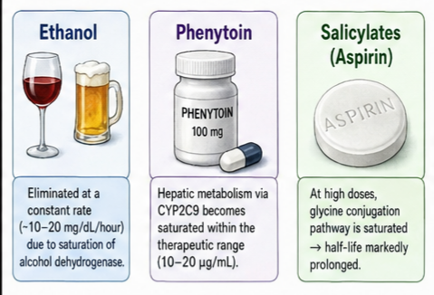
Once alcohol dehydrogenase is saturated, ethanol elimination proceeds at a relatively constant rate in a given individual. The typical elimination rate ranges from 10 to 20 mg per deciliter per hour in alcohol-naive individuals, with an average of approximately 15 mg per deciliter per hour. This rate can increase substantially in chronic heavy drinkers due to enzyme induction, with some individuals eliminating ethanol at rates exceeding 30 mg per deciliter per hour. The zero-order kinetics of ethanol mean that a person presenting with a blood alcohol concentration of 300 mg per deciliter will require approximately 15 to 20 hours to eliminate the alcohol completely, assuming the average elimination rate.
The clinical implications of ethanol’s zero-order elimination are substantial. The duration of intoxication cannot be shortened by any pharmacological intervention; coffee, cold showers, and other folk remedies are ineffective because they do not increase the metabolic capacity of alcohol dehydrogenase. The predictable elimination rate allows clinicians to estimate when a patient will be medically safe for discharge or when their blood alcohol will fall below legal driving limits. In emergency settings, knowledge of ethanol kinetics informs decisions about observation periods, repeat measurements, and the timing of psychiatric or social interventions that require sobriety.
Chronic alcohol consumption produces adaptive changes that alter ethanol kinetics. Induction of CYP2E1 increases the contribution of the microsomal ethanol-oxidizing system, potentially increasing the overall elimination rate. However, liver disease resulting from chronic alcohol use can reduce metabolic capacity, decreasing Vmax and prolonging elimination. The net effect in an individual patient depends on the balance between induction and hepatocellular damage, which must be assessed clinically and through laboratory testing.
Phenytoin: Clinical Application of Michaelis-Menten Kinetics
Phenytoin exemplifies the clinical application of Michaelis-Menten kinetics to therapeutic drug management, and its pharmacokinetic behavior has shaped modern approaches to anticonvulsant therapy and therapeutic drug monitoring. The recognition that phenytoin exhibits saturable metabolism within its therapeutic range has made it one of the most pharmacokinetically challenging drugs in common clinical use, requiring a sophisticated understanding of nonlinear kinetics for safe and effective prescribing.
Phenytoin is eliminated primarily through hepatic metabolism, with the rate-limiting step being para-hydroxylation catalyzed predominantly by CYP2C9, with a minor contribution from CYP2C19. This hydroxylation pathway exhibits saturable kinetics at phenytoin concentrations encountered during routine therapy. The typical Michaelis constant for phenytoin hydroxylation ranges from approximately 4 to 8 mg per liter, which lies near or below the lower end of the commonly cited therapeutic range of 10 to 20 mg per liter. Consequently, phenytoin displays significant nonlinearity throughout its therapeutic range, with the degree of saturation increasing progressively as concentrations rise from the low to high therapeutic range.
The maximum velocity for phenytoin metabolism varies substantially among individuals due to genetic polymorphisms in CYP2C9, concomitant medications that inhibit or induce this isoform, and hepatic function. Typical Vmax values range from approximately 5 to 10 mg per kilogram per day, though wider variation is observed clinically. The interplay between Vmax and the Michaelis constant determines the specific dose-concentration relationship for an individual patient, explaining why some patients achieve therapeutic concentrations with low doses while others require substantially higher doses to reach the same concentration.
The clinical consequence of phenytoin’s saturable metabolism is that dose adjustments must be approached with particular caution, especially as concentrations approach or exceed the upper limit of the therapeutic range. When the serum concentration is low relative to the patient’s Michaelis constant, the relationship between dose and concentration is nearly linear, and standard dose adjustments produce predictable concentration changes. As the concentration rises toward and beyond the Michaelis constant, the dose-concentration curve steepens dramatically. A dose increase of 30 to 50 mg per day, which might produce a concentration increase of 2 to 3 mg per liter at low concentrations, could produce an increase of 5 to 10 mg per liter or more at concentrations in the upper therapeutic range.
Therapeutic drug monitoring for phenytoin requires interpretation of measured concentrations in the context of the patient’s dose and clinical status. Population-based dosing nomograms exist, but their accuracy is limited by interindividual variability in Vmax and Michaelis constant. Bayesian pharmacokinetic approaches, which incorporate population priors with individual patient concentration measurements, provide more accurate dose predictions and are increasingly available through clinical pharmacokinetic software. When interpreting phenytoin concentrations, clinicians must also account for protein binding, as phenytoin is highly bound to albumin, and conditions that reduce albumin concentration or alter binding will change the relationship between total and free drug concentrations.
The management of phenytoin toxicity requires an understanding that elimination will follow zero-order kinetics when concentrations are substantially elevated. At toxic concentrations, the elimination rate is essentially constant at Vmax, and the time required for concentrations to return to the therapeutic range can be estimated by dividing the excess concentration by the zero-order rate constant. Extracorporeal removal is generally ineffective for phenytoin because of its high protein binding and large volume of distribution, so management consists primarily of supportive care while awaiting metabolic elimination.
High-Dose Aspirin and Salicylate Kinetics
Aspirin, or acetylsalicylic acid, undergoes rapid deacetylation to salicylic acid, which is responsible for both the anti-inflammatory effects and the dose-dependent pharmacokinetics that produce zero-order elimination at high concentrations. The kinetics of salicylate elimination illustrate the importance of understanding metabolic pathway saturation in the context of both therapeutic anti-inflammatory dosing and the management of acute and chronic salicylate toxicity.
Salicylate elimination occurs through multiple parallel pathways, each with different kinetic characteristics. At low doses producing salicylate concentrations below approximately 15 to 20 mg per deciliter, elimination follows first-order kinetics with a half-life of approximately 2 to 4 hours. The primary elimination routes at these concentrations include renal excretion of unchanged salicylate and formation of salicyluric acid through conjugation with glycine, catalyzed by the enzyme glycine N-acyltransferase. Additional pathways include formation of salicylphenolic glucuronide and salicylacyl glucuronide, as well as oxidation to gentisic acid.
At higher salicylate concentrations, the glycine conjugation pathway becomes saturated because the availability of glycine and the capacity of glycine N-acyltransferase are limited. When this occurs, the elimination pattern shifts from first-order toward zero-order kinetics, with the half-life extending to 15 to 30 hours or longer at concentrations exceeding 30 to 40 mg per deciliter. This prolongation of half-life has profound implications for both therapeutic dosing and toxicity. Patients receiving high-dose aspirin for inflammatory conditions must be monitored carefully, as small dose increases can produce disproportionate increases in serum salicylate concentrations once the metabolic pathways approach saturation.
The clinical manifestations of salicylate toxicity reflect both the direct effects of salicylates on multiple organ systems and the metabolic derangements resulting from uncoupling of oxidative phosphorylation. Mild toxicity, with serum concentrations of 30 to 50 mg per deciliter, may produce tinnitus, nausea, and hyperventilation. Moderate toxicity, at concentrations of 50 to 80 mg per deciliter, can cause fever, metabolic acidosis with respiratory alkalosis, and altered mental status. Severe toxicity, with concentrations exceeding 80 to 100 mg per deciliter, may result in coma, seizures, cardiovascular collapse, and death. The zero-order elimination kinetics at high concentrations mean that toxicity can be prolonged, with resolution requiring many hours to days depending on the peak concentration achieved.
The management of salicylate toxicity incorporates pharmacokinetic principles in several ways. Urinary alkalinization with intravenous sodium bicarbonate enhances renal salicylate elimination by ion trapping, increasing the clearance of salicylate independent of the saturable metabolic pathways. Hemodialysis effectively removes salicylate and is indicated for severe toxicity, particularly when concentrations exceed 80 to 100 mg per deciliter or when end-organ toxicity is present. The decision to initiate hemodialysis must account for the expected duration of toxicity based on zero-order elimination kinetics, as prolonged exposure to high salicylate concentrations increases the risk of irreversible organ damage.
Additional Drugs Exhibiting Zero-Order Kinetics
Beyond ethanol, phenytoin, and salicylates, several additional clinically important drugs exhibit zero-order or saturable elimination kinetics under specific circumstances. Familiarity with these agents allows clinicians to anticipate nonlinear pharmacokinetic behavior and adjust prescribing and monitoring practices accordingly.
Theophylline, a methylxanthine bronchodilator used in the management of asthma and chronic obstructive pulmonary disease, demonstrates dose-dependent elimination kinetics that can become clinically significant at concentrations near or above the upper limit of the therapeutic range. Theophylline is metabolized primarily through hepatic CYP1A2, with a minor contribution from renal excretion. At therapeutic concentrations of 5 to 15 mg per liter, elimination typically follows first-order kinetics in adults, with a half-life of 6 to 9 hours. However, at concentrations exceeding 20 to 25 mg per liter, the metabolic pathways begin to saturate, and the elimination pattern shifts toward zero-order kinetics. This transition means that patients with theophylline toxicity, whether from acute overdose or chronic accumulation, may have prolonged elimination half-lives extending to 20 hours or more. The clinical consequences include sustained toxicity requiring extended monitoring and, in severe cases, consideration of extracorporeal removal with hemodialysis or hemoperfusion.
Voriconazole, a triazole antifungal agent, exhibits nonlinear pharmacokinetics due to saturable metabolism by CYP2C19, with additional contributions from CYP3A4 and CYP2C9. The saturable nature of voriconazole metabolism results in disproportionate increases in plasma concentration with dose escalation, complicating the achievement of therapeutic concentrations while avoiding toxicity. The nonlinear kinetics, combined with genetic polymorphisms in CYP2C19 that create distinct poor, intermediate, extensive, and ultrarapid metabolizer phenotypes, produce wide interindividual variability in voriconazole exposure. Therapeutic drug monitoring is recommended to guide voriconazole dosing and ensure concentrations within the therapeutic range, particularly given the established relationship between voriconazole concentrations and both efficacy and toxicity.
Fluorouracil, an antimetabolite chemotherapeutic agent, undergoes saturable metabolism through dihydropyrimidine dehydrogenase, the rate-limiting enzyme in pyrimidine catabolism. The capacity of this enzyme determines the clearance of fluorouracil, and saturation at higher doses produces nonlinear pharmacokinetics. Patients with partial or complete dihydropyrimidine dehydrogenase deficiency, resulting from genetic polymorphisms, are at risk for severe and potentially life-threatening toxicity when exposed to standard doses of fluorouracil because their already-limited metabolic capacity is more readily saturated. Pretreatment screening for dihydropyrimidine dehydrogenase deficiency and pharmacokinetic-guided dosing are emerging strategies to improve the safety of fluorouracil therapy.
Heparin demonstrates zero-order elimination characteristics at high doses due to saturation of both the reticuloendothelial clearance system and the endothelial binding sites that mediate its removal from the circulation. At low doses, heparin elimination involves a rapid saturable phase representing binding to endothelial cells and macrophages, followed by slower renal elimination. At therapeutic anticoagulant doses, the saturable phase becomes saturated, and elimination transitions toward first-order kinetics, with dose-dependent half-lives that complicate the transition between heparin and other anticoagulants.
Factors Affecting Zero-Order Elimination Kinetics
The parameters governing zero-order elimination vary substantially among individuals and within the same individual over time, reflecting the influence of genetic, physiological, pathological, and pharmacological factors on drug-metabolizing capacity. Understanding these sources of variability is essential for predicting individual patient responses to drugs with saturable metabolism and for interpreting unexpected changes in drug concentrations.
Genetic polymorphisms in drug-metabolizing enzymes represent a major source of interindividual variability in zero-order elimination parameters. The cytochrome P450 enzymes exhibit extensive genetic variation, with polymorphisms affecting both the quantity and catalytic activity of expressed enzyme. For phenytoin, variants in CYP2C9, particularly the CYP2C9*2 and CYP2C9*3 alleles, reduce enzymatic activity and lower Vmax, increasing the tendency toward saturation at lower drug concentrations. Patients carrying these variant alleles require lower phenytoin doses to achieve therapeutic concentrations and are at increased risk for concentration-dependent toxicity. Similarly, CYP2C19 polymorphisms influence voriconazole metabolism, with poor metabolizers exhibiting higher concentrations and greater nonlinearity at standard doses compared to extensive metabolizers.
Age-related changes in hepatic function and enzyme expression affect the capacity for drug metabolism and, consequently, the parameters of zero-order elimination. Neonates and infants have immature drug-metabolizing enzyme systems with reduced Vmax values compared to adults, increasing their susceptibility to saturation kinetics. Elderly patients experience progressive reductions in liver mass, hepatic blood flow, and enzyme activity, all of which can reduce Vmax and alter the concentration at which saturation occurs. Age-related changes in protein binding and renal function add additional complexity, potentially affecting both the free drug concentration available for metabolism and the contribution of renal elimination to overall clearance.
Hepatic disease reduces the metabolic capacity of the liver through loss of functional hepatocytes, altered hepatic architecture, and diminished blood flow. For drugs metabolized primarily through hepatic pathways, liver disease can substantially reduce Vmax and lower the concentration threshold at which saturation occurs. The Child-Pugh classification, which incorporates clinical and laboratory markers of hepatic dysfunction, provides a framework for estimating the degree of metabolic impairment, though individual variation in residual enzyme function remains substantial. In patients with significant hepatic disease, drugs with saturable metabolism should be initiated at lower doses and titrated more cautiously, with therapeutic drug monitoring employed where available.
Drug interactions affecting zero-order kinetics occur when concomitant medications inhibit or induce the enzymes responsible for the saturable metabolic pathway. Enzyme inhibition reduces Vmax, effectively lowering the ceiling on elimination capacity and causing saturation at lower drug concentrations. Potent CYP2C9 inhibitors such as fluconazole and amiodarone can substantially reduce phenytoin clearance, precipitating toxicity in patients previously stable on their phenytoin regimen. Enzyme induction increases Vmax, raising the capacity for drug elimination and reducing the tendency toward saturation. Rifampin, a broad-spectrum inducer of multiple cytochrome P450 enzymes, can increase the Vmax for phenytoin metabolism, requiring higher doses to maintain therapeutic concentrations.
Toxicity Risks Associated with Zero-Order Kinetics
The toxicity risks inherent in zero-order drug elimination stem directly from the loss of the homeostatic relationship between concentration and elimination rate that characterizes first-order kinetics. When elimination capacity is saturated, the normal mechanisms that prevent excessive drug accumulation are disabled, and concentrations can escalate rapidly with continued drug administration or with minor changes in metabolic capacity.
The most significant toxicity risk is the nonlinear relationship between dose and steady-state concentration. For drugs with zero-order kinetics at therapeutic concentrations, the margin between a dose that produces therapeutic effects and a dose that produces toxicity can be narrow and difficult to predict based on dosing alone. A patient maintained on a stable phenytoin regimen may develop toxicity when a small dose increase is prescribed, when a new medication inhibits phenytoin metabolism, or when hepatic function declines due to intercurrent illness. The absence of a constant half-life means that toxic concentrations will persist longer than expected based on experience with first-order drugs, as the elimination system cannot accelerate to clear the excess drug.
Drug accumulation represents another toxicity mechanism for zero-order agents. When the daily dose of a drug exceeds the maximum elimination capacity, steady state is never achieved, and drug concentrations continue to rise indefinitely until toxicity intervenes or the dose is reduced. This scenario can develop insidiously with drugs that have long half-lives at therapeutic concentrations but transition to saturation kinetics with chronic dosing. Clinicians must be vigilant for signs of accumulation, including escalating serum concentrations on serial monitoring and the emergence of concentration-dependent adverse effects.
The variability in metabolic capacity among individuals creates a population-level toxicity risk, as standard dosing recommendations derived from pharmacokinetic studies in healthy volunteers or selected patient populations may not apply to individuals with reduced enzyme activity due to genetics, age, disease, or concomitant medications. A phenytoin dose that produces therapeutic concentrations in an extensive CYP2C9 metabolizer may cause toxicity in a poor metabolizer, even though both patients appear similar based on demographic characteristics. This variability underscores the importance of individualized dosing and therapeutic drug monitoring for zero-order drugs.
The prolonged duration of toxicity following overdose of zero-order drugs poses challenges for clinical management. Supportive care must be maintained for extended periods while awaiting metabolic elimination, consuming healthcare resources and exposing patients to the risks of prolonged hospitalization. The decision to employ extracorporeal removal techniques such as hemodialysis must balance the risks of the procedure against the expected duration of spontaneous elimination, a calculation that depends directly on zero-order kinetic parameters.
Therapeutic Drug Monitoring for Zero-Order Drugs
Therapeutic drug monitoring provides the clinical tool necessary to manage the pharmacokinetic variability and nonlinearity inherent in zero-order drug elimination. By measuring drug concentrations in individual patients, clinicians can individualize dosing to achieve therapeutic targets while avoiding concentrations associated with toxicity. The principles and practice of therapeutic drug monitoring for zero-order drugs differ in important ways from monitoring for first-order agents.
The rationale for therapeutic drug monitoring rests on the relationship between drug concentration and clinical effect. For drugs with established therapeutic ranges, maintaining concentrations within these ranges maximizes the probability of therapeutic response while minimizing the risk of toxicity. This rationale is strengthened for zero-order drugs, where the relationship between dose and concentration is unpredictable and where concentration-dependent toxicity is a significant clinical concern. For phenytoin, numerous studies have demonstrated improved seizure control and reduced toxicity when dosing is guided by serum concentration monitoring compared to empiric dose adjustment.
The timing of sample collection for therapeutic drug monitoring must account for the kinetic behavior of the drug being measured. For drugs with saturable metabolism, trough concentrations measured immediately before the next dose provide the most consistent measure of drug exposure and the most reliable basis for dose adjustment. Peak concentrations may be measured when acute toxicity is suspected following intravenous loading doses or when absorption kinetics are in question. The interpretation of measured concentrations requires knowledge of the time since the last dose, the dosing interval, and the expected concentration-time profile based on the drug’s kinetic characteristics.
The interpretation of measured drug concentrations for zero-order drugs requires a different conceptual framework than for first-order agents. For first-order drugs, the relationship between dose change and concentration change is linear and proportional: doubling the dose doubles the concentration. This proportionality allows straightforward dose adjustments based on the ratio of the desired concentration to the measured concentration. For zero-order drugs, the same proportional adjustment will overestimate the required dose change at higher concentrations and underestimate it at lower concentrations. Instead, Bayesian pharmacokinetic approaches that incorporate the Michaelis-Menten model with population-based prior estimates of Vmax and Michaelis constant, updated with individual patient concentration measurements, provide more accurate dose predictions.
Practical aspects of therapeutic drug monitoring include the selection of appropriate assay methods, the establishment of therapeutic ranges based on clinical outcome data, and the integration of concentration data with clinical assessment. Immunoassay methods provide rapid turnaround for commonly monitored drugs such as phenytoin, while chromatographic methods offer greater specificity when metabolites or co-administered drugs might interfere. The therapeutic range must be understood as a probabilistic guide rather than an absolute boundary, with some patients achieving therapeutic effects at concentrations below the range and others requiring concentrations above it.
Dose Adjustment Principles for Saturation Kinetics
Adjusting doses for drugs that exhibit zero-order or Michaelis-Menten elimination kinetics requires an approach distinct from the proportional adjustments appropriate for first-order agents. The principles governing dose adjustment for saturation kinetics derive from the mathematical properties of the Michaelis-Menten equation and have been refined through clinical experience with phenytoin, theophylline, and other drugs exhibiting dose-dependent elimination.
The fundamental principle is that the dose change required to produce a desired concentration change depends on the current concentration relative to the patient’s Michaelis constant. At low concentrations, where the system operates in the approximately linear portion of the Michaelis-Menten curve, dose adjustments can be made proportionally. A measured concentration of 5 mg per liter with a target of 10 mg per liter might reasonably be achieved by doubling the dose, assuming the Michaelis constant is substantially above this range. At higher concentrations approaching or exceeding the Michaelis constant, proportional adjustments become inaccurate and dangerous, as the same proportional increase would produce a much larger concentration change than anticipated.
The clinical approach to dose adjustment involves estimating the patient’s individual pharmacokinetic parameters from available concentration data and then using these parameters to calculate the dose expected to achieve the target concentration. At minimum, two steady-state concentration measurements obtained at different dose levels are required to solve the Michaelis-Menten equation for Vmax and Michaelis constant. With these parameters estimated, the dose needed to achieve any target concentration can be calculated. Bayesian methods enhance this approach by incorporating population-based prior distributions for the pharmacokinetic parameters, weighted according to the precision of the individual patient’s data.
For practical clinical purposes, simpler approaches have been developed and validated. For phenytoin, the concept of the effective Vmax, representing the maximum daily elimination at the current concentration, can be used to estimate the dose change needed for small concentration adjustments. The effective Vmax is calculated from the current dose and concentration using a rearrangement of the Michaelis-Menten equation that assumes a population-average Michaelis constant. While less accurate than methods using individually estimated parameters, this approach provides clinically useful dose guidance when only a single concentration measurement is available.
The cardinal rule for dose adjustment with zero-order drugs is to make changes in small increments, particularly when the current concentration is in the upper portion of the therapeutic range or above it. For phenytoin, dose changes of 30 mg per day, or approximately 10 percent of a typical maintenance dose, are recommended when concentrations are near or above 15 mg per liter. Larger changes may be appropriate when concentrations are clearly subtherapeutic and the patient is far from saturation. The response to any dose change should be assessed with repeat concentration measurement after a suitable interval, acknowledging that the time to reach a new steady state will be longer at higher concentrations due to the prolonged half-life associated with saturation.
Clinical Case Scenarios Illustrating Zero-Order Kinetics
The application of zero-order kinetic principles to clinical practice is best illustrated through representative case scenarios that demonstrate how these concepts inform diagnostic reasoning, therapeutic decision-making, and patient management. The following cases, while fictionalized, reflect common clinical situations encountered in emergency medicine, neurology, and primary care.
Case One: Ethanol Intoxication in the Emergency Department
A 38-year-old man is brought to the emergency department by paramedics after being found confused and unsteady in a public park. On arrival, he is drowsy but arousable, with slurred speech, horizontal nystagmus, and an ataxic gait. His vital signs are notable for mild tachycardia at 102 beats per minute. A serum ethanol concentration returns at 280 mg per deciliter. The emergency physician must determine the appropriate disposition and timing.
Applying zero-order kinetic principles, the physician estimates that ethanol elimination proceeds at approximately 15 mg per deciliter per hour in this patient, who has no history of chronic alcohol use that might induce accelerated metabolism. The concentration will reach 100 mg per deciliter, a level below which most non-tolerant individuals show minimal intoxication, in approximately 12 hours. The concentration will reach levels acceptable for safe discharge from a cognitive standpoint in approximately 15 to 18 hours. The physician orders repeat ethanol measurements at 6 and 12 hours to confirm the predicted decline and to document that concentrations are falling as expected, ruling out ongoing absorption from a gastric ethanol reservoir.
This case illustrates the clinical utility of zero-order kinetics for predicting the duration of intoxication, planning appropriate monitoring intervals, and determining medically appropriate discharge timing. The constant rate of elimination, while inconvenient for the patient and the emergency department, provides a reliable basis for these predictions.
Case Two: Phenytoin Toxicity Following Dose Adjustment
A 52-year-old woman with epilepsy secondary to a previous traumatic brain injury has been maintained on phenytoin 300 mg daily for three years, with serum concentrations consistently in the range of 14 to 17 mg per liter. She has been seizure-free on this regimen. After experiencing a breakthrough seizure, her neurologist increases the phenytoin dose to 400 mg daily. Two weeks later, she presents to clinic complaining of dizziness, unsteadiness, and blurred vision. A serum phenytoin concentration returns at 34 mg per liter.
The patient’s symptoms are consistent with phenytoin neurotoxicity, and the concentration is well above the therapeutic range. The dose increase of 100 mg per day, representing a 33 percent increase, produced a near-doubling of the serum concentration because she was already operating in the nonlinear portion of the Michaelis-Menten curve at her baseline concentration. The neurologist reduces the dose to 330 mg daily and arranges for repeat concentration measurement in two weeks, anticipating that this more cautious increment will achieve a concentration in the low 20s mg per liter, providing enhanced seizure protection without crossing into the toxic range.
This case demonstrates the dangers of applying proportional dose adjustments to drugs with saturable kinetics and illustrates the importance of small, incremental dose changes guided by concentration monitoring when operating near the saturation point.
Case Three: Salicylate Toxicity from Chronic Dosing
A 68-year-old man with osteoarthritis has been self-medicating with over-the-counter aspirin for joint pain, gradually escalating his dose as his symptoms have worsened. He presents to his primary care physician with a two-week history of tinnitus, subjective hearing loss, and intermittent confusion. His wife reports that he has been breathing more rapidly than usual and has seemed intermittently confused. A serum salicylate concentration returns at 48 mg per deciliter, and blood gas analysis reveals a mixed metabolic acidosis and respiratory alkalosis.
The patient’s presentation is classic for chronic salicylate toxicity, also known as salicylism. His gradual dose escalation has allowed salicylate to accumulate as the glycine conjugation pathway became saturated. At his current concentration, elimination is proceeding primarily through zero-order kinetics, with a half-life of approximately 20 hours. The physician discontinues aspirin therapy, initiates intravenous fluids with sodium bicarbonate to alkalinize the urine and enhance renal salicylate excretion, and arranges for hospitalization with serial salicylate concentration monitoring. The patient improves over 48 hours as concentrations decline through the combined effects of metabolic and enhanced renal elimination.
This case illustrates how saturation kinetics can produce insidious toxicity with gradual dose escalation, the characteristic clinical features of salicylism, and the management principles that incorporate both supportive care and interventions to enhance elimination independent of the saturated metabolic pathways.
Examination Questions for Medical Students and Residents
The principles of zero-order kinetics appear frequently in pharmacology, internal medicine, and emergency medicine examinations. The following representative questions, with detailed explanations, illustrate the types of knowledge assessment that medical students and residents should be prepared to address.
Question 1
A 45-year-old man presents to the emergency department with ethanol intoxication. His initial serum ethanol concentration is 300 mg/dL. Assuming an average elimination rate of 15 mg/dL per hour, approximately how long will it take for his serum ethanol concentration to decrease to 60 mg/dL?
A. 4 hours
B. 8 hours
C. 16 hours
D. 24 hours
E. 32 hours
Answer: C. 16 hours
Explanation: Ethanol follows zero-order elimination kinetics at the concentrations achieved during intoxication. The elimination rate is constant at approximately 15 mg/dL per hour. The total concentration decrease required is 300 minus 60, equaling 240 mg/dL. Dividing the required decrease by the elimination rate gives 240 divided by 15, equaling 16 hours. Note that the time would be the same if the initial concentration were 200 mg/dL and the target were 140 mg/dL, because the same absolute decrease is required. This illustrates the fundamental difference between zero-order kinetics, where elimination time depends on the absolute concentration change, and first-order kinetics, where elimination time would depend on the fractional concentration change.
Question 2
Which of the following statements best describes the elimination kinetics of phenytoin at therapeutic concentrations?
A. Phenytoin follows first-order kinetics with a constant half-life of 22 hours
B. Phenytoin follows zero-order kinetics with a constant elimination rate of 5 mg/kg/day
C. Phenytoin follows Michaelis-Menten kinetics with dose-dependent half-life
D. Phenytoin elimination is independent of hepatic enzyme activity
E. Phenytoin elimination increases proportionally with increasing dose
Answer: C. Phenytoin follows Michaelis-Menten kinetics with dose-dependent half-life
Explanation: Phenytoin is eliminated through saturable hepatic metabolism following Michaelis-Menten kinetics. At therapeutic concentrations, which are near or above the typical Michaelis constant, phenytoin displays characteristics of both first-order and zero-order elimination, and its half-life is concentration-dependent. Option A is incorrect because phenytoin does not have a constant half-life; the half-life increases at higher concentrations. Option B is incorrect because while phenytoin can approach zero-order kinetics at high concentrations, it is not purely zero-order throughout the therapeutic range. Option D is incorrect because phenytoin elimination is heavily dependent on CYP2C9 activity. Option E describes first-order kinetics, which does not apply to phenytoin at therapeutic concentrations.
Question 3
A patient with epilepsy has been maintained on phenytoin 300 mg daily with a steady-state serum concentration of 12 mg/L. The neurologist increases the dose to 400 mg daily. Two weeks later, the serum concentration is 26 mg/L. Which of the following best explains this disproportionate increase in concentration?
A. First-order elimination with reduced clearance
B. Enzyme induction by the higher phenytoin dose
C. Saturation of hepatic metabolic pathways
D. Increased bioavailability of the higher dose
E. Laboratory error in one of the measurements
Answer: C. Saturation of hepatic metabolic pathways
Explanation: The 33 percent increase in daily dose produced a 117 percent increase in serum concentration, indicating nonlinear pharmacokinetics consistent with saturation of the CYP2C9-mediated metabolic pathway. At a baseline concentration of 12 mg/L, the patient was already operating above the typical Michaelis constant for phenytoin, in a region where the dose-concentration relationship is nonlinear. The additional dose exceeded the remaining metabolic capacity more than would be predicted by linear kinetics. Option A would produce a proportional increase. Option B would increase metabolism, producing lower concentrations. Option D is unlikely for a well-absorbed drug. Option E should be considered but does not explain the consistent pattern of nonlinear kinetics well-established for phenytoin.
Question . What is zero-order kinetics in simple terms?
Question . Why is ethanol considered the classic example of zero-order kinetics?
Question . Can a drug follow both first-order and zero-order kinetics?
Question . How does zero-order elimination affect drug dosing?
Question . What is the relationship between Michaelis-Menten kinetics and zero-order kinetics?
Question . How does therapeutic drug monitoring help manage zero-order drugs?
Question . Why do some drugs have a narrow therapeutic index due to zero-order kinetics?
Question . What factors can increase the risk of toxicity with zero-order drugs?
Question . How does the management of overdose differ for zero-order drugs?
Question . Are there any clinical advantages to zero-order drug elimination?
Question . How does enzyme induction affect zero-order kinetics?
Question . What is the clinical significance of zero-order kinetics for medical students to understand?
Key Takeaways
Zero-order kinetics represents a critical concept in clinical pharmacology with direct implications for drug safety, therapeutic monitoring, and patient care. The following key points summarize the essential information presented in this review.
The defining characteristic of zero-order elimination is a constant rate of drug removal that remains independent of drug concentration. This occurs when the enzymatic or transport systems responsible for elimination become saturated, operating at their maximum capacity. Drugs that exhibit zero-order kinetics lack a constant half-life, instead displaying concentration-dependent elimination times that complicate dosing and monitoring.
The Michaelis-Menten model provides the theoretical framework for understanding saturable elimination kinetics, describing the transition from first-order behavior at low concentrations to zero-order behavior when metabolic systems are saturated. The two key parameters of this model are Vmax, the maximum elimination rate, and the Michaelis constant, the concentration at which elimination proceeds at half the maximum rate.
Ethanol, phenytoin, high-dose salicylates, and several other clinically important drugs exhibit zero-order kinetics under circumstances commonly encountered in clinical practice. For ethanol, saturation of alcohol dehydrogenase occurs at concentrations achieved after minimal alcohol consumption, producing the characteristic constant elimination rate of 10 to 20 mg/dL per hour. Phenytoin displays Michaelis-Menten kinetics throughout its therapeutic range, with the degree of nonlinearity increasing as concentrations rise, requiring cautious dose adjustment and therapeutic drug monitoring.
The clinical implications of zero-order kinetics include unpredictable dose-concentration relationships, narrow therapeutic indices, prolonged toxicity following overdose, and the essential role of therapeutic drug monitoring in guiding therapy. Dose adjustments for zero-order drugs must be made in small increments, particularly when concentrations are in the upper therapeutic range or above, and should be guided by measured serum concentrations whenever possible.
Understanding the factors that affect zero-order elimination parameters, including genetic polymorphisms, age, hepatic disease, and drug interactions, allows clinicians to anticipate individual variability in drug response and to adjust prescribing accordingly. The principles of zero-order kinetics extend beyond the specific drugs discussed in this review to inform the broader understanding of pharmacokinetic principles and their application to patient care.
Conclusion
Zero-order elimination kinetics represents far more than an abstract pharmacological concept confined to textbooks and examination questions. It embodies a fundamental biological principle with immediate and practical consequences for patient care across multiple medical specialties. The saturation of drug-metabolizing systems, with its attendant loss of the protective proportionality between concentration and elimination rate, creates clinical challenges that demand respect, understanding, and vigilance from prescribing clinicians.
The drugs that exhibit zero-order kinetics include some of the oldest and most widely used agents in the pharmacopeia, as well as newer medications for which saturable metabolism was recognized during clinical development. Ethanol, consumed by a substantial fraction of the global population and responsible for enormous morbidity and healthcare utilization, follows zero-order kinetics that determine the duration of intoxication and inform emergency management. Phenytoin, a mainstay of anticonvulsant therapy for decades, requires nuanced understanding of its Michaelis-Menten kinetics for safe long-term use. Salicylates, available without prescription and frequently implicated in both intentional and unintentional poisonings, exhibit dose-dependent elimination that transforms therapeutic agents into dangerous toxins when metabolic pathways are overwhelmed.
The recognition that zero-order kinetics arises from the finite capacity of biological systems carries implications beyond the specific drugs discussed in this review. The principle that enzymatic processes can become saturated applies broadly in pharmacology, from drug metabolism to receptor binding to transport processes. Clinicians who understand saturation kinetics are better equipped to anticipate drug interactions that occur through competition for limited metabolic capacity, to recognize the potential for nonlinear dose-response relationships with new medications, and to appreciate the importance of therapeutic drug monitoring for drugs with narrow therapeutic indices.
For medical students and healthcare professionals in training, mastery of zero-order kinetics provides a foundation for understanding more complex pharmacokinetic concepts and their clinical applications. The ability to recognize which drugs exhibit saturation kinetics, to predict how saturation will affect dosing and toxicity, and to apply therapeutic drug monitoring principles appropriately distinguishes the competent clinician from the merely knowledgeable. As pharmacology education continues to emphasize the integration of basic science principles with clinical practice, zero-order kinetics serves as an exemplary topic that bridges the gap between biochemical mechanisms and bedside decision-making.
The safe and effective use of medications requires not only knowledge of indications, contraindications, and adverse effects but also an understanding of the pharmacokinetic principles that determine drug disposition in individual patients. Zero-order kinetics, while representing a minority of therapeutic agents, accounts for a disproportionate share of drug-related toxicity and therapeutic failures. By applying the principles detailed in this review, clinicians can minimize the risks associated with saturation kinetics while maximizing the therapeutic benefits of drugs that exhibit this challenging but manageable pharmacokinetic behavior.
References
Benet LZ, Hoener BA. Changes in plasma protein binding have little clinical relevance. Clinical Pharmacology and Therapeutics. 2002;71(3):115-121.
Browne TR, Kugler AR, Eldon MA. Pharmacology and pharmacokinetics of fosphenytoin. Neurology. 1996;46(6 Suppl 1):S3-S7.
Brunton LL, Hilal-Dandan R, Knollmann BC, editors. Goodman and Gilman’s The Pharmacological Basis of Therapeutics. 14th ed. New York: McGraw-Hill Education; 2023.
Gibaldi M, Perrier D. Pharmacokinetics. 2nd ed. New York: Marcel Dekker; 1982.
Katzung BG, Vanderah TW, editors. Basic and Clinical Pharmacology. 16th ed. New York: McGraw-Hill Education; 2023.
Levy G. Pharmacokinetics of salicylate elimination in man. Journal of Pharmaceutical Sciences. 1965;54(7):959-967.
Ludden TM. Nonlinear pharmacokinetics: clinical implications. Clinical Pharmacokinetics. 1991;20(6):429-446.
Michaelis L, Menten ML. Die Kinetik der Invertinwirkung. Biochemische Zeitschrift. 1913;49:333-369.
Mullen W, Laskin OL. The kinetics of ethanol elimination in man. Journal of Pharmacology and Experimental Therapeutics. 1982;220(1):151-156.
Patsalos PN, Berry DJ, Bourgeois BF, et al. Antiepileptic drugs—best practice guidelines for therapeutic drug monitoring: a position paper by the subcommission on therapeutic drug monitoring, ILAE Commission on Therapeutic Strategies. Epilepsia. 2008;49(7):1239-1276.
Richens A, Dunlop A. Serum phenytoin levels in management of epilepsy. The Lancet. 1975;306(7928):247-248.
Ritchie JM, editor. The Pharmacological Basis of Therapeutics. 5th ed. New York: Macmillan; 1975.
Rowland M, Tozer TN. Clinical Pharmacokinetics: Concepts and Applications. 4th ed. Philadelphia: Lippincott Williams and Wilkins; 2011.
Temple AR. Acute and chronic effects of aspirin toxicity and their treatment. Archives of Internal Medicine. 1981;141(3 Spec No):364-369.
Thomson AH, Whiting B. Bayesian parameter estimation and population pharmacokinetics. Clinical Pharmacokinetics. 1992;22(6):447-467.
Tozer TN, Winter ME. Phenytoin. In: Evans WE, Schentag JJ, Jusko WJ, editors. Applied Pharmacokinetics: Principles of Therapeutic Drug Monitoring. 3rd ed. Vancouver: Applied Therapeutics; 1992.
U.S. Food and Drug Administration. Guidance for Industry: Population Pharmacokinetics. Rockville, MD: FDA; 2022.
U.S. Food and Drug Administration. Phenytoin Sodium Prescribing Information. Available from FDA Online Label Repository.
U.S. Food and Drug Administration. Voriconazole Prescribing Information. Available from FDA Online Label Repository.
Widmark EMP. Principles and Applications of Medicolegal Alcohol Determination. Davis, CA: Biomedical Publications; 1981.
Winter ME. Basic Clinical Pharmacokinetics. 5th ed. Philadelphia: Lippincott Williams and Wilkins; 2010.
World Health Organization. Guidance for the Safe and Effective Use of Antiepileptic Drugs. Geneva: WHO Press; 2020.
Yasir M, Goyal A, Sonthalia S. Corticosteroid Adverse Effects. In: StatPearls. Treasure Island, FL: StatPearls Publishing; 2023.
Zandvliet AS, Huitema AD, Copalu W, et al. CYP2C9 and CYP2C19 polymorphic forms are related to increased indisulam exposure and higher hematologic toxicity. Clinical Cancer Research. 2007;13(10):2970-2976.
Disclaimer: This article is intended for educational and informational purposes only. It does not constitute medical advice. Always consult a qualified healthcare professional for diagnosis and treatment of medical conditions.